
|
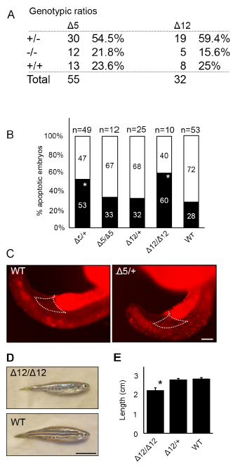
Fig. S5
Hemogen mutant zebrafish have increased cell death during embryonic development.
(A) Genotypic ratios of 2 dpf embryos produced from heterozygous incrosses of Hemgnnuz3 (Δ5) or Hemgnnuz4 (Δ12) mutants. (B) Proportion of genotyped mutants and wild-type sibling embryos at 2 dpf that were apoptotic (black) or phenotypically normal (white) (* P ≤ 0.05, ** P ≤ 0.005, Chi square). (BC) Acridine orange staining for apoptotic cells is increased in the bodies and in the peripheral blood island (outlined) in 20 hpf heterozygous Hemgnnuz2 mutant zebrafish (n = 3) compared to wild-type siblings (n = 3). (D) Comparison of adult wild-type and homozygous Hemgnnuz4 (Δ12) mutant. (E) Average body length of adult wild-type and Hemgnnuz4 (Δ12) mutants. Error bars represent standard error (*, P ≤ 0.05, Student’s t test). Scale bars = 100 μm (E), 50 mm (D)