
|
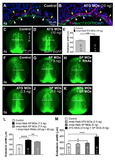
Fig. S7
Mmp14a/b are required for C&E movements of the anterior endodermal cells.
(A-B) Confocal z-stack images of transverse cryosections from Tg(sox17:EGFP) control embryos and embryos injected with mmp14a/b ATG MOs (10ng, suppression of translation) immunostained for Fn (magenta) and nuclei (DAPI, blue). Fn assembly at mes/end (white arrowheads) and ect/mes (yellow arrowheads) boundaries. (C-M) Embryos injected with indicated MOs targeting mmp14a/b (ATG MOs target the translation; SP MOs target the splicing). (C-D, F-K) Epifluorescence still images of the anterior region of the endodermal sheet in the indicated embryos. Anterior-dorsal view. A, anterior; P, posterior. White lines of equivalent length indicate the width of anterior endodermal sheets of the embryos at the same stage. (E) Average endodermal width at the anterior region of embryos shown in (C,D). (L) Average width of anterior endoderm in embryos shown in (F-H). (M) Average width of anterior endoderm in embryos shown in (I-K). The number of embryos analyzed in each group is indicated. **, p<0.01; ***, p<0.001; ****, p<0.0001; #, p>0.05, student’s t-test.