
|
Fig. S5
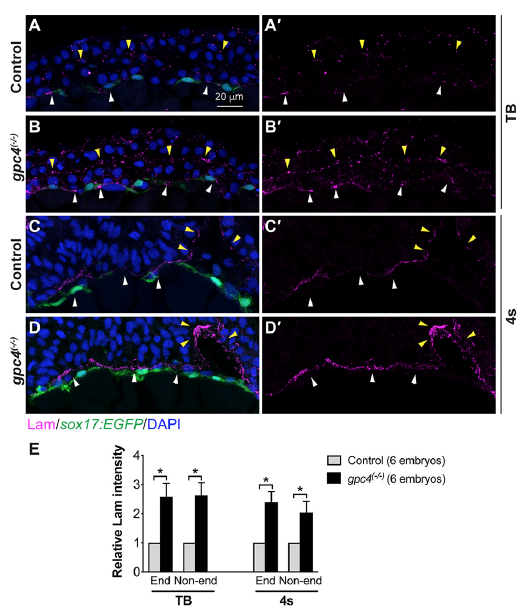
Lam deposition is increased in gpc4 mutant embryos.
Transverse cryosections from Tg(sox17:EGFP) control and gpc4 mutant embryos immunostained for Lam (magenta) and nuclei (DAPI, blue). (A-D) Confocal z-stack images of embryos at tailbud (TB) (AB ) and 4-somite (4s) (C-D) stages. Lam assembly between the ectoderm and mesoderm (yellow arrowheads) and around the endodermal layer (white arrowheads). (E) Relative Lam intensity in nonendodermal (Non-end) tissue and around the endodermal layer (End) in control and gpc4 mutant embryos at TB and 4s. The number of embryo analyzed is shown in the graph. Bars represent the mean±s.e.m. *, P<0.05, student’s t-test.