
|
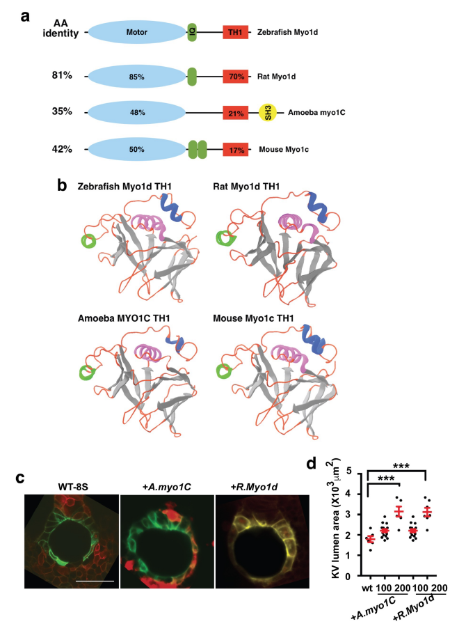
Fig. S5
Amino acid comparison of myosin1 showing protein domains.
a. Class I myosins showing myosin motor (blue), calmodulin binding IQ motif (green), Tail Homology 1 (TH1, red) and SH3 (yellow) domains. Total amino acid identity is listed on the left column compared to zebrafish Myo1d. Amino acid identity to zebrafish Myo1D motor and TH1 motifs are listed within the domains.
b. Images of the predicted TH1 domains showing similarity after Phyre2 modeling.
c. Representative images of KVs from Tg(dusp6:GFP-MA) embryos injected with H2BmCherry mRNA and A. myo1C, or Rat Myo1d showing expansion of the KV lumen area.
d. Graph showing KV lumen area after injection of various doses of myosin I family. Statistical comparisons for the graphs were by one-way ANOVA and post hoc analysis with Turkey’s multiple range tests. *** p<0.001 represents a statistical difference Data shown in the graphs are mean ± SEM.