
Image
|
Figure Caption
Fig. S2
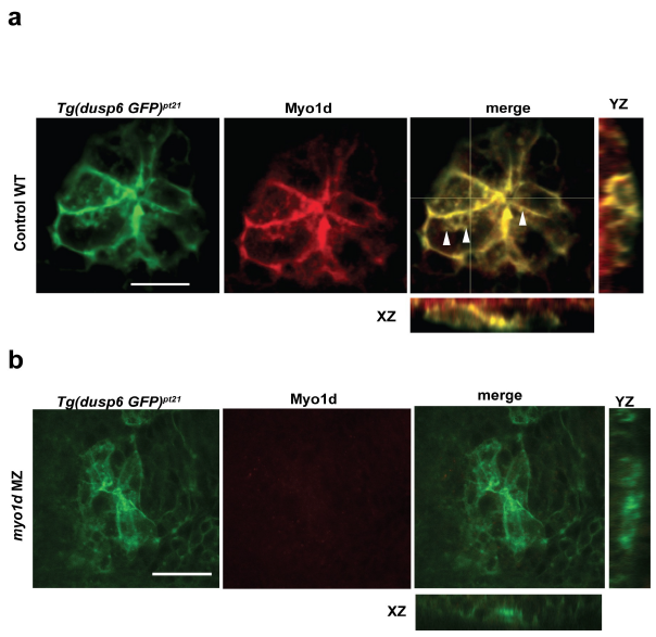
Myo1D expression in KV epithelial cells colocalizes with vacuoles and are absent in myo1d MZ mutants.
a. Tg(dusp6:GFP-MA) embryos stained with Myo1D antibody (red) shows co-localized expression of Myo1D to membranes, and punctate structures in KV epithelial cells at 1-2 S.
b. Myo1d staining in myo1d MZ embryos shows loss of Myo1D expression, suggesting the pt31a allele is a null mutant (scale bar-50μm).
Acknowledgments
This image is the copyrighted work of the attributed author or publisher, and
ZFIN has permission only to display this image to its users.
Additional permissions should be obtained from the applicable author or publisher of the image.
Full text @ Nat. Commun.