
Image
|
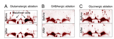
Figure Caption
Fig. S5
Ablation of neurotransmitter-specified Gsx1 neurons dorsal to Mauthner cell. Related to Figure 5
A-C. Glutamatergic (A: gsx1-Gal4, UAS:Cre-ERT2, vglut2a:Switch-GFP, red), GABAergic (B: gsx1-Gal4, UAS:Cre-ERT2, gad1b:Switch-GFP, red), and glycinergic (C: gsx1:Cre, glyt2:Switch-Gal4,UAS:GFP, red) Gsx1 neurons in the dorsal vicinity of the Mauthner cell (retrogradely labeled, red) before (top) and after (bottom) laser ablation. Scale bar 50 μm applies to all panels.
Acknowledgments
This image is the copyrighted work of the attributed author or publisher, and
ZFIN has permission only to display this image to its users.
Additional permissions should be obtained from the applicable author or publisher of the image.
Full text @ Curr. Biol.