
Fig. 4
- ID
- ZDB-IMAGE-180921-2
- Genes
- Publication
- Song et al., 2018 - Developmental expression of the zebrafish Arf-like small GTPase paralogs arl13a and arl13b
- All Figures
- Figures for Song et al., 2018
|
Fig. 4
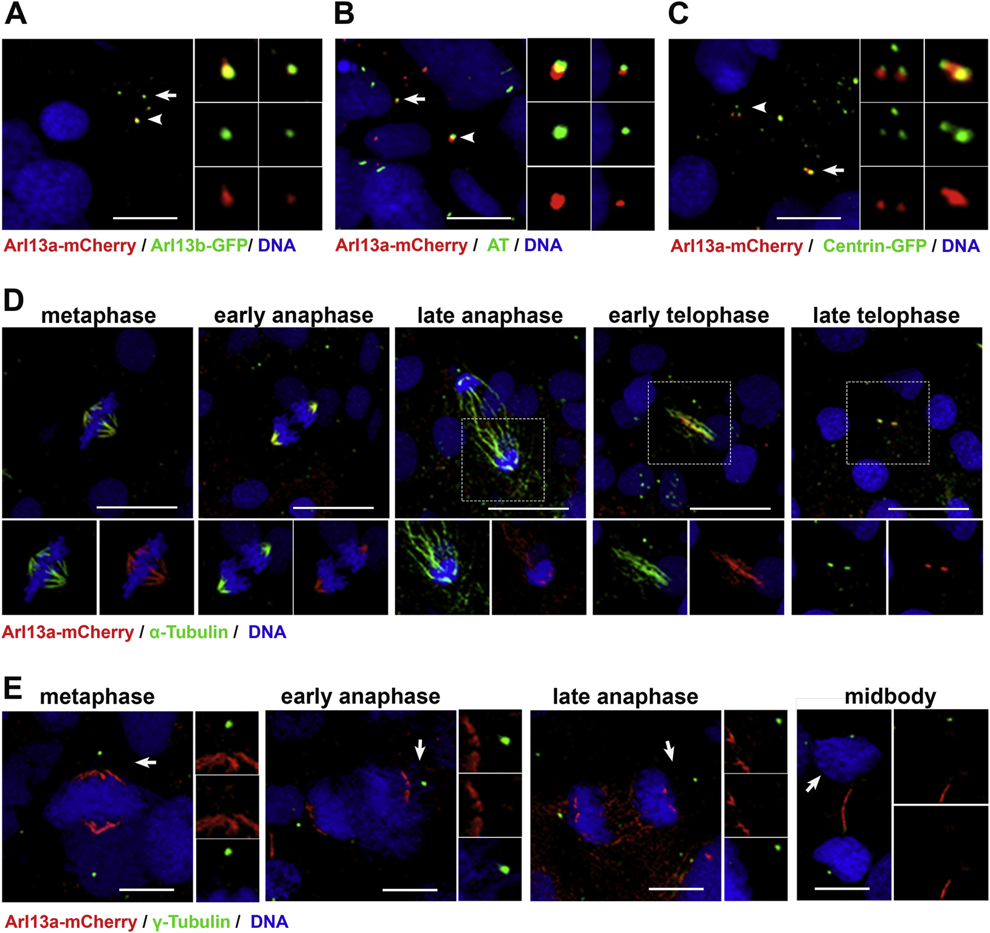
Arl13a colocalizes with microtubules in zebrafish embryos. (A) Tg(-3.2actnb:arl13b-GFP) transgenic larvae injected with mRNA encoding Arl13a-mCherry were fixed at 6 somite stage (ss) and stained with antibodies against mCherry (red) or GFP (green). Cilia on two cells (arrow and arrowhead). Inset panels show enlarged images of individual channels and the merged image. (B) Wild-type larvae (6 ss) were fixed and stained with antibodies against mCherry (red) or acetylated-tubulin (green) to label cilia. (C) Tg(-3.2actnb:cetn4-GFP) transgenic larvae injected with mRNA encoding Arl13a-mCherry were fixed at 6 somite stage (ss) and stained with antibodies against mCherry (red) or GFP (green). (D) Mitotic cells in presomitic mesoderm of wild-type larvae (6 ss) were fixed and stained with antibodies against mCherry (red) or acetylated-tubulin (green) to label microtubules or (E) γ-tubulin to label centrosomes. All larvae were counterstained with DAPI to label nuclei. Scale bar: 10 μm (A–C); 20 μm (D, E).
Reprinted from Gene expression patterns : GEP, 29, Song, P., Perkins, B.D., Developmental expression of the zebrafish Arf-like small GTPase paralogs arl13a and arl13b, 82-87, Copyright (2018) with permission from Elsevier. Full text @ Gene Expr. Patterns