
Image
|
Figure Caption
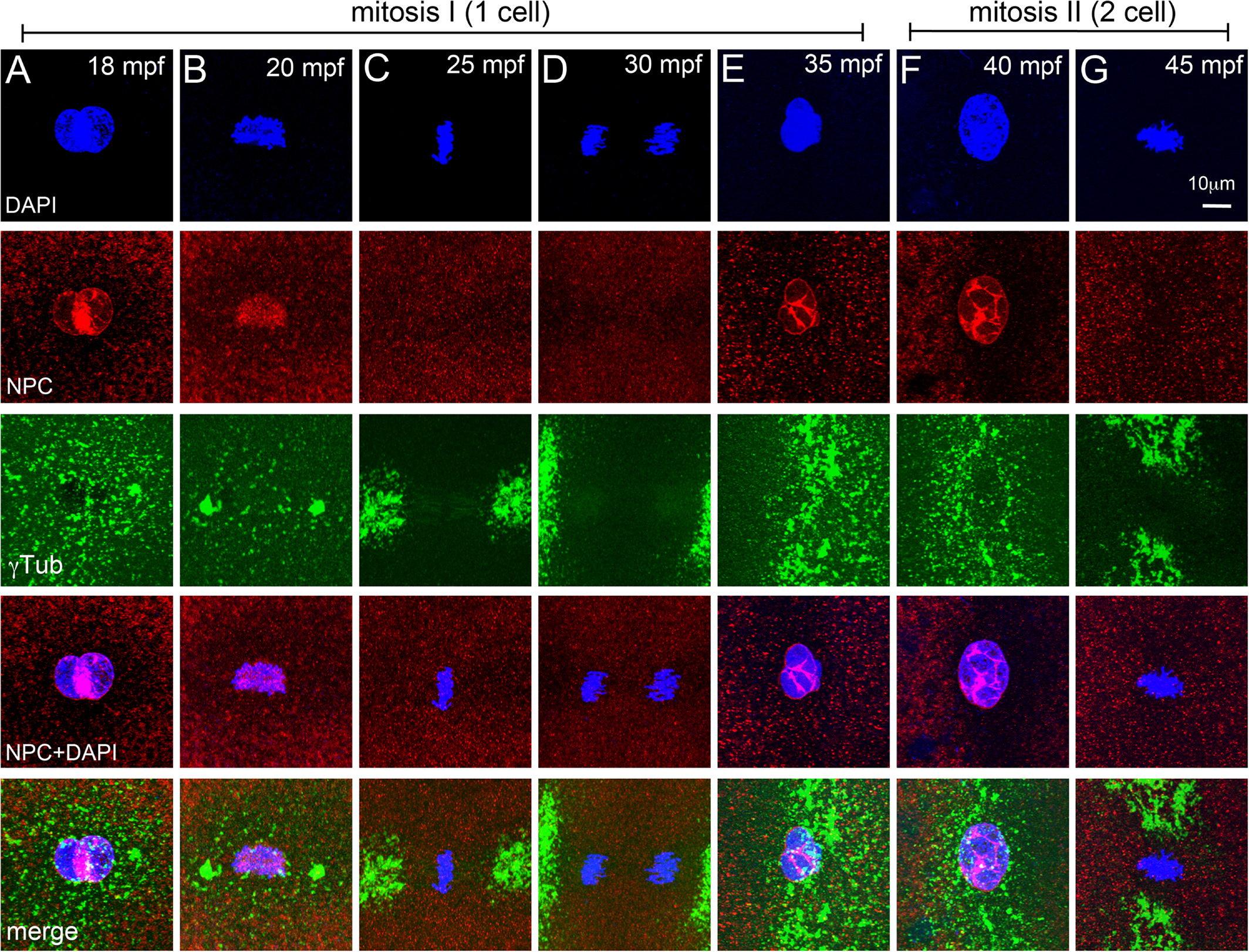
Fig. 7
NEBD cycles during early mitosis in zebrafish embryos. A–G: Immunofluorescence labelings of control embryos for DNA (DAPI, blue), NPC (red), and γ‐tubulin (green). Nuclear envelope exists around pronuclei (A), disassembles at onset of and during mitosis‐I (B–D), re‐forms during telophase‐I (E), and exists during prophase‐II (F), but disassembles again for mitosis‐II (G). In panels E–G only one of the two cells is shown. Each panel is a representative image of 4 to 6 embryos imaged for a particular time point.
Acknowledgments
This image is the copyrighted work of the attributed author or publisher, and
ZFIN has permission only to display this image to its users.
Additional permissions should be obtained from the applicable author or publisher of the image.
Full text @ Dev. Dyn.