
|
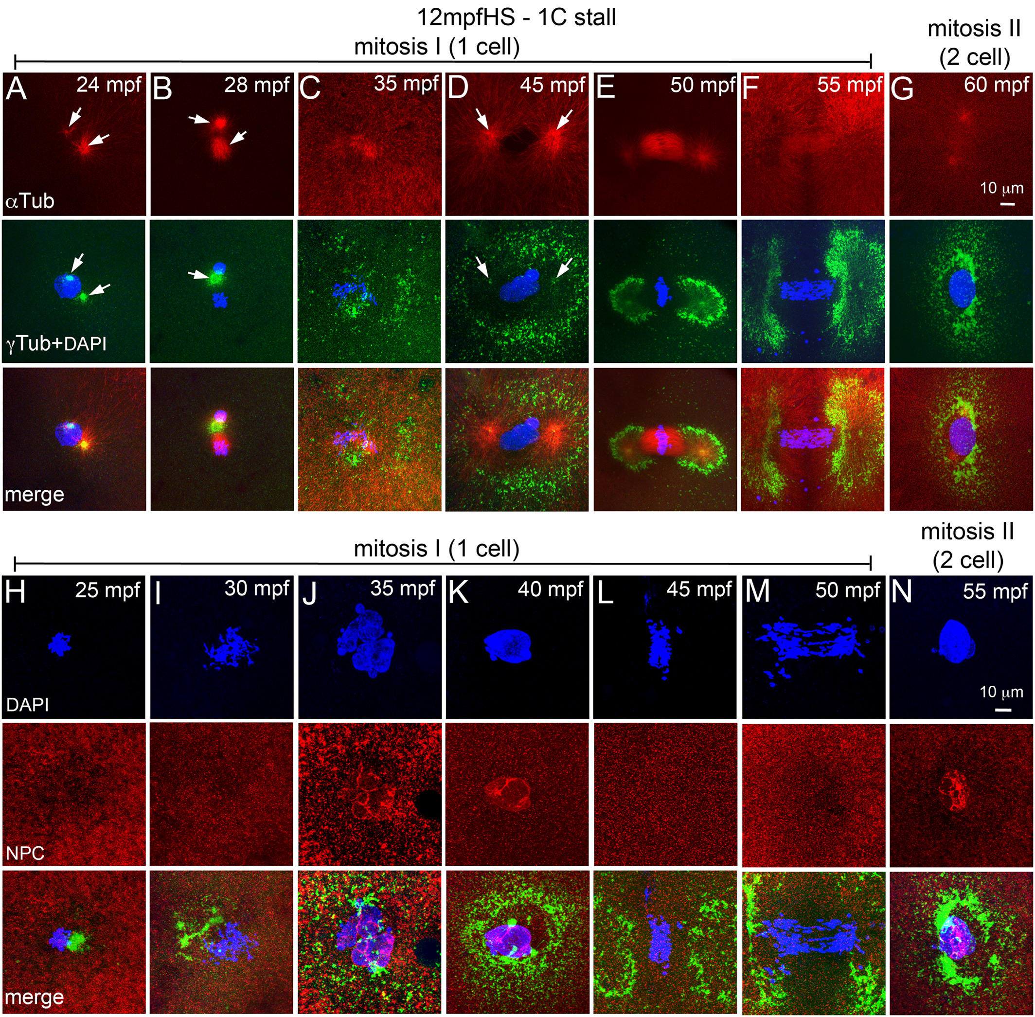
Fig. 4
12 mpfHS delays entry into mitosis‐I and does not affect NEBD. A–G: Immunofluorescence labeling of 12 mpfHS embryos for DNA (DAPI, blue), α‐tubulin (red) and γ‐tubulin (green). Centrosomes are mispositioned (A,B), PCM recovers (C), and mitosis‐I proceeds with prophase‐I (D), metaphase‐I (E), anaphase‐I (F), and telophase‐I (G). Arrows indicate microtubule asters in the α‐tubulin panels and centrosomal foci in the γ‐tubulin panels. H–N: Immunofluorescence labeling of 12 mpfHS embryos for DNA (DAPI, blue), NPC (red), and γ‐tubulin (green). Nuclear envelope does not exist after heat shock at 12 mpf (H,I), is re‐formed at ∼35 mpf (J,K), breaks down upon entry into mitosis‐I (L,M), and re‐forms at the end of mitosis‐I (N). In panels G and N only one of the two cells is shown. For panels A–G, each panel is a representative image of 10 to 15 embryos imaged for a particular time point. For panels H–N, each panel is a representative image of 4 to 6 embryos imaged for a particular time point.