
Image
|
Figure Caption
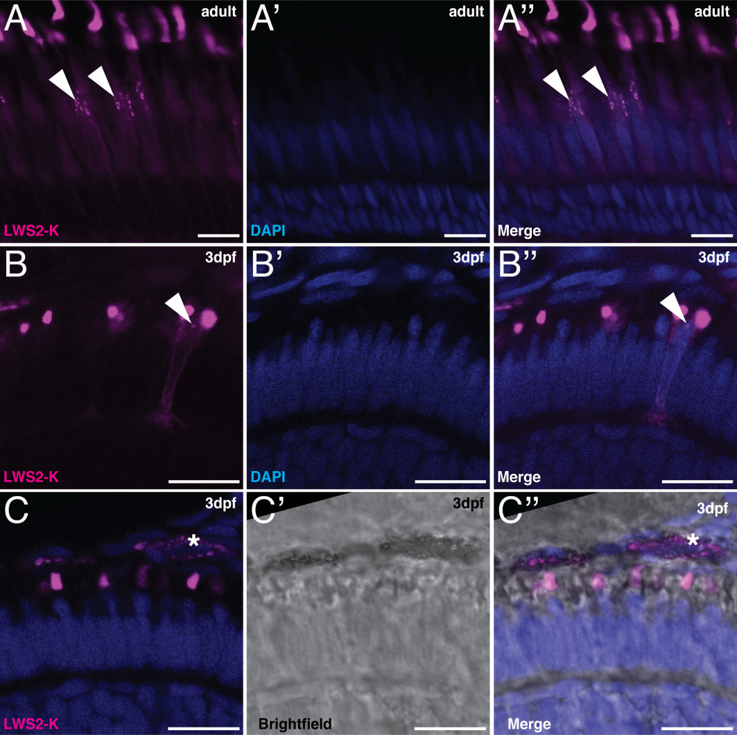
Fig. 4
LWS2‐K signals in embryonic and adult retinas. Retinal sections of Tg(LWS) with LWS2‐K in magenta and DAPI in blue. A–B″: Vesicle‐like structures are present in the inner segment of LWS2‐K–positive cells. A–A″: Retinal sections of adult Tg(LWS). B–B″: Retinal sections of Tg(LWS) at 3 dpf. C–C″: Retinal sections of Tg(LWS) with brightfield image in gray scale. Asterisks indicate the LWS2‐K–positive signal in the retinal pigment epithelium, which is likely due to the pigment present in these cells. Scale bars A–C″ = 10 μm.
Acknowledgments
This image is the copyrighted work of the attributed author or publisher, and
ZFIN has permission only to display this image to its users.
Additional permissions should be obtained from the applicable author or publisher of the image.
Full text @ Dev. Dyn.