
Image
|
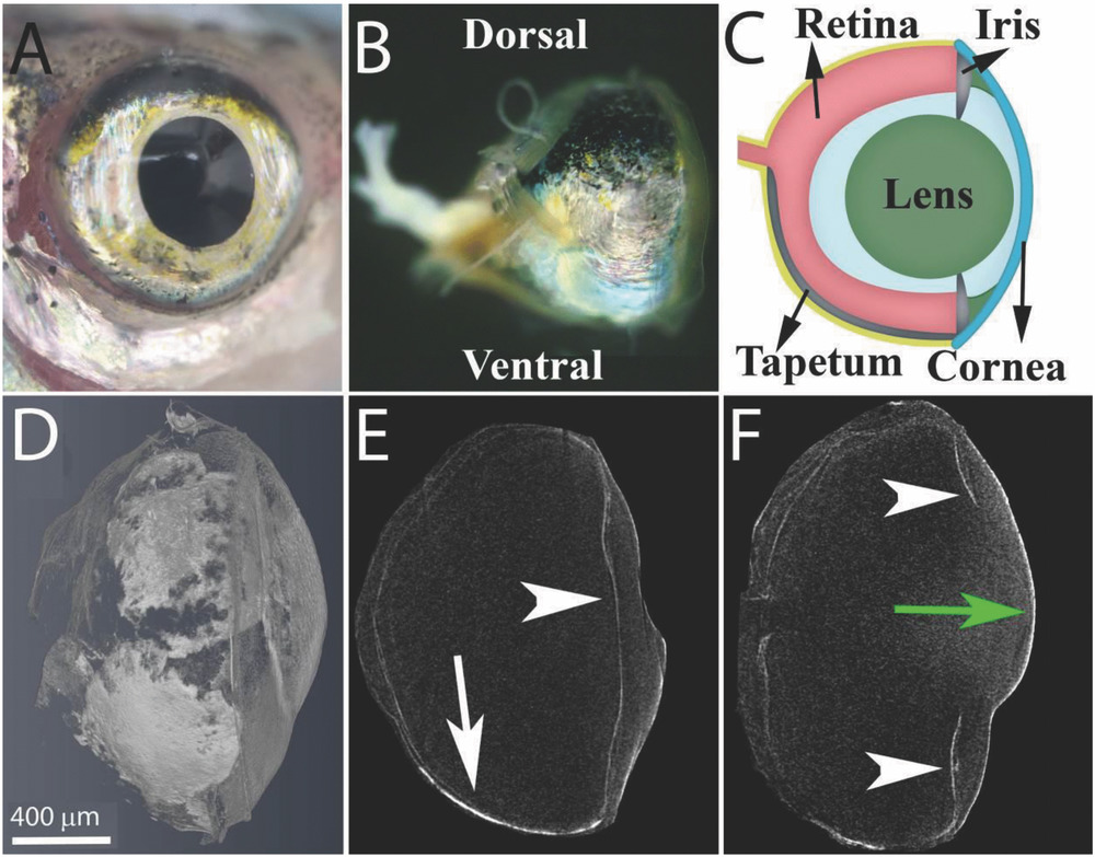
Figure Caption
Fig. 1
The anatomy of the zebrafish eye. A) A top view of the zebrafish eye showing the silvery iris. B) A side view of the fish eye showing the tapetum lucidum, a silvery layer, located at the ventral side of the eyeball and a pigmented layer visible at the dorsal side of the eye. C) A schematic representation of the eye showing the cornea (blue), lens (green), and retina (light red), as well as the guanine‐based reflecting layers, the iris argenta and the tapetum lucidum (silver). D–F) Micro‐CT images of the zebrafish eye: a volume rendering of the whole eye, a sagittal view of the lower part of the eye, and a sagittal view of the center of the eye. Arrowheads indicate the iris argentum, white arrow indicates the tapetum lucidum, and green arrow indicates the cornea. Scale bars: 400 µm.
Acknowledgments
This image is the copyrighted work of the attributed author or publisher, and
ZFIN has permission only to display this image to its users.
Additional permissions should be obtained from the applicable author or publisher of the image.
Full text @ Adv Sci (Weinh)