
Image
|
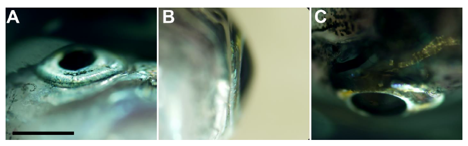
Figure Caption
Fig. S6
Light microscope images of the outer iridophores surrounding the eye.
This layer extends to the point where the eye is protruding out from the fish head. A) Ventral view of the eye. B) A side view from posterior to anterior. C) A dorsal view of the eye.
Acknowledgments
This image is the copyrighted work of the attributed author or publisher, and
ZFIN has permission only to display this image to its users.
Additional permissions should be obtained from the applicable author or publisher of the image.
Full text @ Adv Sci (Weinh)