
Image
|
Figure Caption
Fig. 5-S1
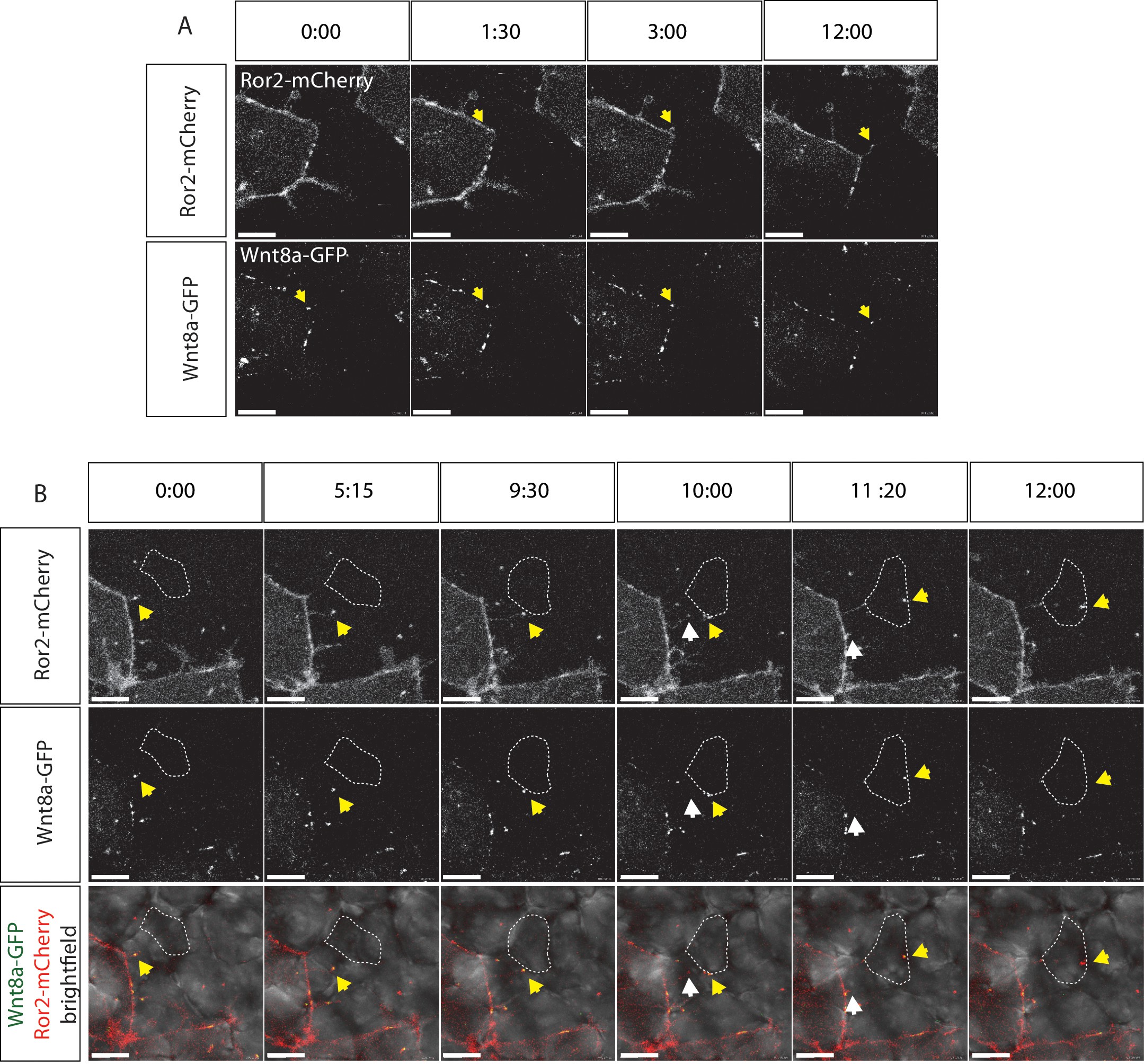
Visualization of Ror2-Wnt8a cytonemal transport in the living zebrafish.
Time series of still pictures of single channels of Ror2-mCherry/Wnt8a-GFP-expressing cells to observe (A) Wnt8a recruitment to the membrane and cytoneme initiation and (B) cytonemal target finding and Ror2/Wnt8a cluster endocytosis into the receiving cell. White arrows indicate the Wnt8a/Ror2 cluster whereas the yellow arrows highlight pruning of the cytoneme tip after successful cytonemal delivery. (B) A series of merged pictures with the bright field channel are included in this panel. Scale bars = 10 µm; in (E) = 20 µm.
Acknowledgments
This image is the copyrighted work of the attributed author or publisher, and
ZFIN has permission only to display this image to its users.
Additional permissions should be obtained from the applicable author or publisher of the image.
Full text @ Elife