
|
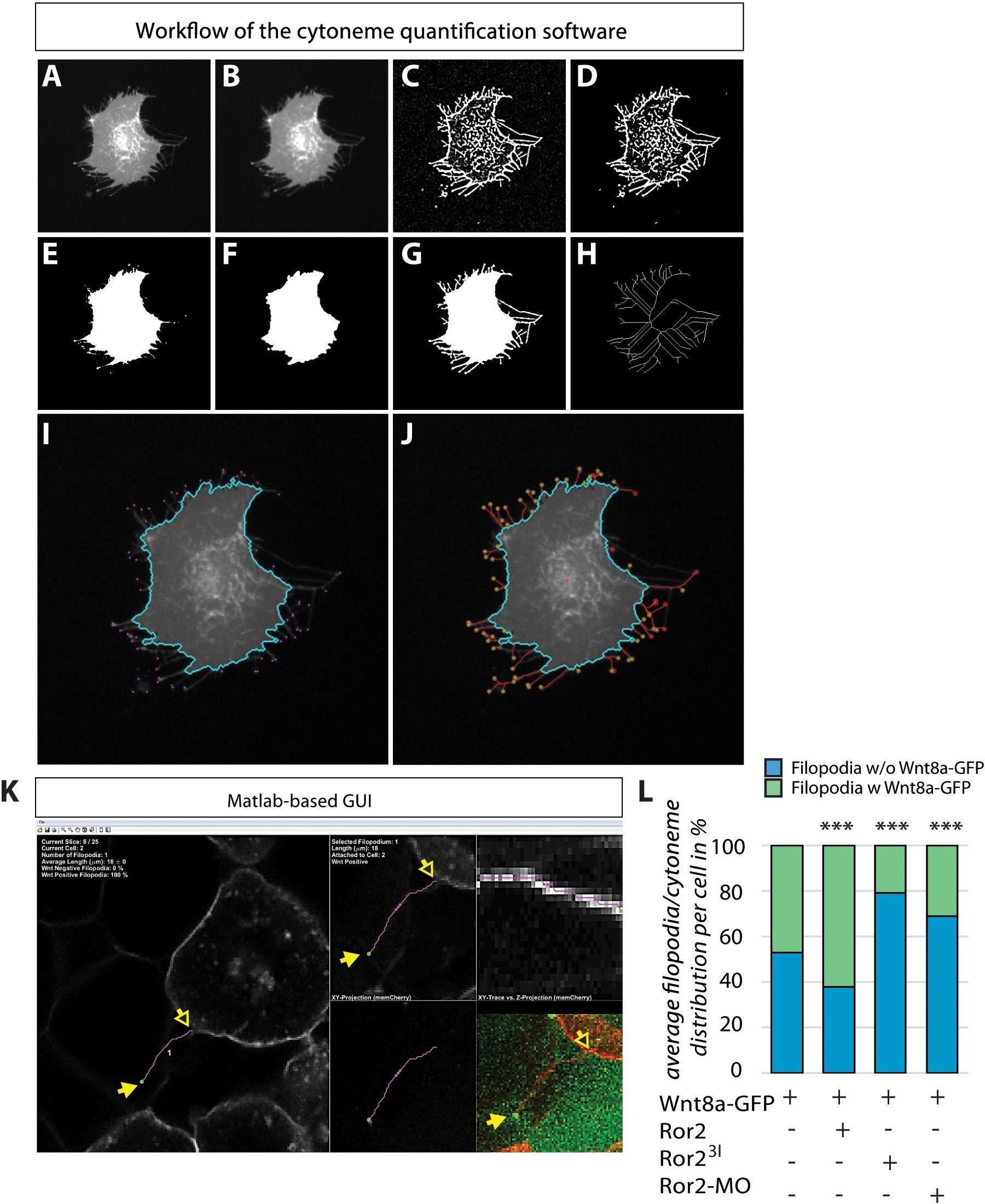
Fig. 4-S1
Cytoneme quantification software toextract filopodia automatically using the memCherry channel of the acquired images.
The raw image (A) was initially smoothed with a Gaussian low-pass filter (B) to reduce image noise for facilitated processing. To emphasize the filopodia in the image, we used an objectness filter that emphasizes line-like structures based on the eigenvalues of the Hessian matrix (C) and that binarized the resulting edge image (D) using local adaptive thresholding. For the detection of the cell body, we applied a local adaptive threshold to the Gaussian smoothed raw image (E) and performed a morphological opening operation to remove filopodia remains from the cell body (F). The two binarized images (D) and (F) were then combined to yield the final segmentation mask including cell body and filopodia (G). Finally, the combined binary image (G) was used to obtain a skeleton image (H), which in turn allows the extraction of potential filopodia tips at the end points of the skeleton. The identified cell body (cyan outline) and the potential filopodia tips (magenta dots) are shown in (I). A livewire approach was then used to automatically trace filopodia from the tips to the cell body (J, red lines). (K) To measure and analyse filopodia in acquired 3D images, we developed a custom-made MATLAB graphical user interface (GUI) that allowed us to scroll through the slices of the stack and to trace filopodia semi-automatically on the basis of the manually provided start and end points of a filopodium of interest (yellow arrows). The software automatically traces the filopodium in 3D using a livewire approach to obtain accurate length quantifications. The software also keeps track of all segmented filopodia, provides filopodia counts and length measurements, and provides average quantifications of all identified filopodia. In addition, in a window of the GFP channel, we were able to distinguish between Wnt-positive and Wnt-negative filopodia for embryos expressing Wnt8a-GFP.