
|
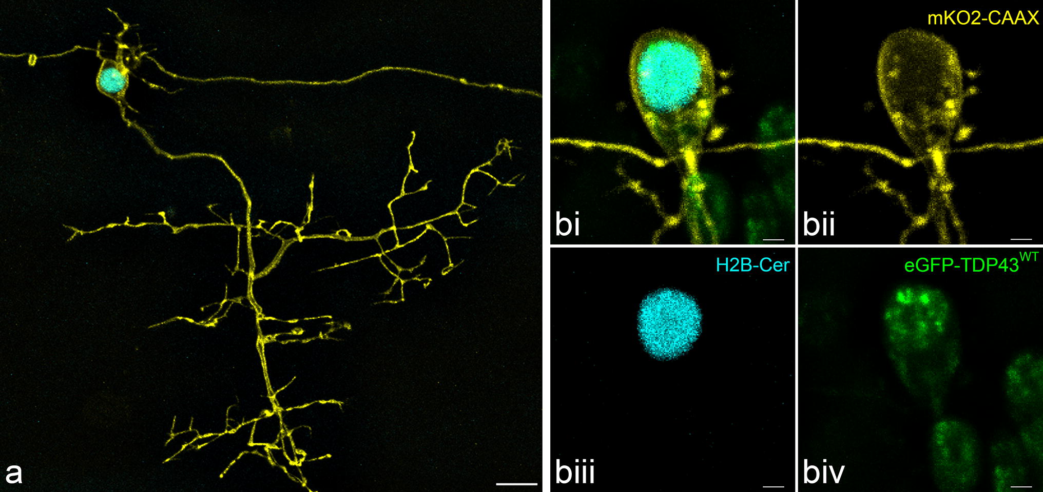
Fig. 1
Fluorescent primary motor neurons in the zebrafish spinal cord at 3 dpf. a Maximum intensity projection of a z-stack of a motor neuron revealing the complete axonal arbour (yellow) projecting into and around the myotome (not shown). At the cell body the spindly dendritic arbour can also be observed. The nucleus is visualised in mCerulean3 (cyan). Traversing the image is the axon of a spinal interneuron. Scale = 10 µm. b A separate fluorescent motor neuron (bi), demonstrating the mnx1-driven membrane-bound mKO2-CAAX (yellow, bii), nuclear H2B-mCerulean3 (cyan, biii) and HsaTDP43WT-linked eGFP (green, biv). Scale = 2 µm