
|
Fig. 3
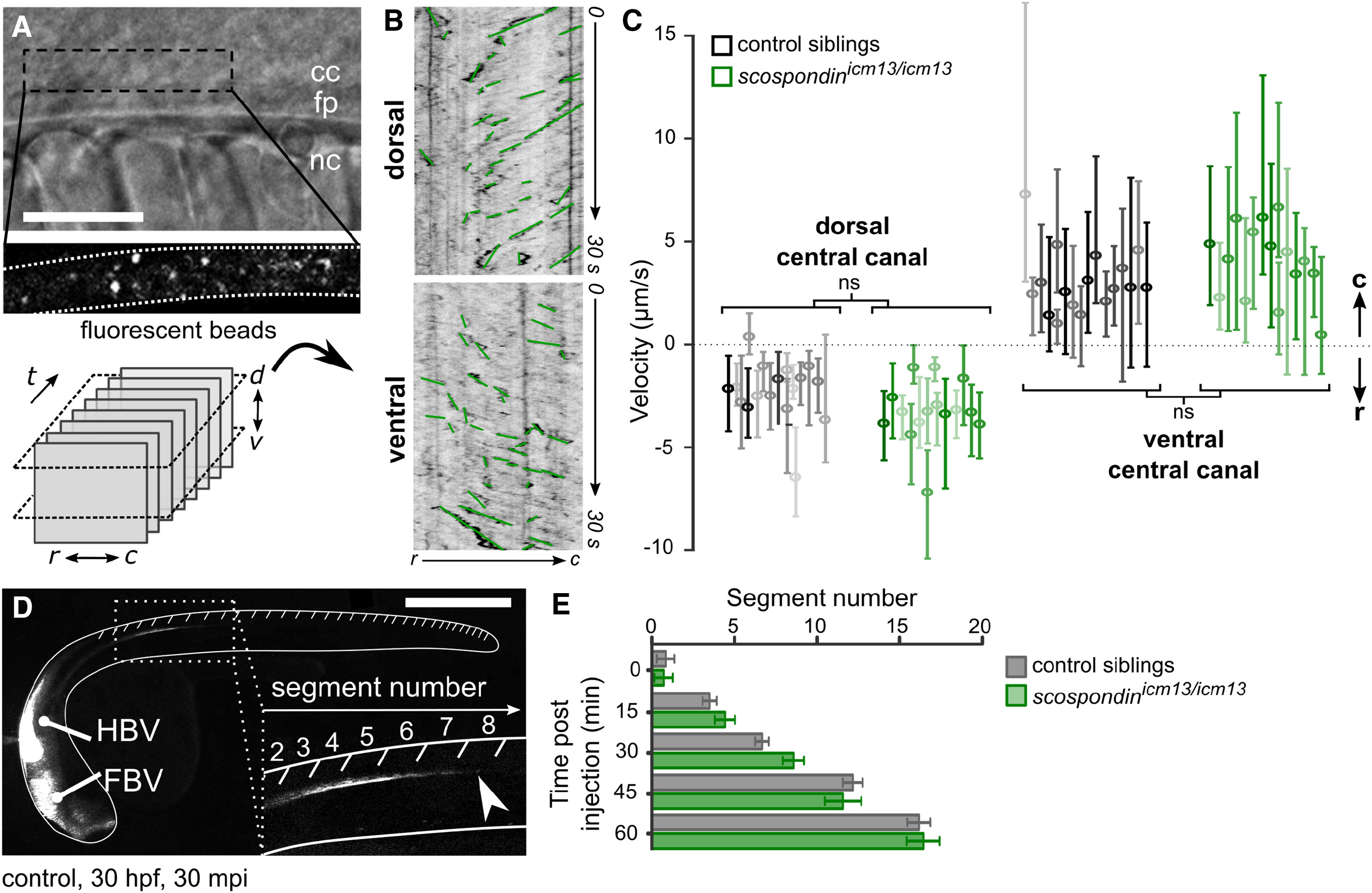
Cerebrospinal Fluid Properly Flows in the Central Canal of the scospondinicm13/icm13 Mutant
(A and B) Lateral view of the central canal (A; transmitted, top) filled with fluorescent beads (bottom). Scale bar represents 30 μm. Time-lapse images at two positions (dorsal and ventral) are represented with the rostro-caudal axis as the horizontal axis and time as the vertical one (B). Kymographs reveal a bidirectional flow, with bead trajectories pointing at opposite directions in the dorsal and ventral central canal. Bead trajectories (green) were used to estimate bead velocities along the rostro-caudal axis.
(C) Bead velocities were similar in control siblings (black) and scospondinicm13/icm13 (green) embryos in the dorsal and ventral central canal (control: n = 570; 584; mutants: n = 582; 634 trajectories in the dorsal; ventral position, respectively; p = 0.07 and 0.33, t = −1.8 and 0.99, and degrees of freedom [df] = 28.8 and 16 in the dorsal and ventral position, respectively, two-tailed t test). Values are given as median ± interquartile range; one boxplot per fish; color intensity reflects the number of measured trajectories per fish. ns, not significant.
(D) 30 hpf zebrafish embryo injected with fluorescent beads in brain ventricles shows transport down the central canal (inset) 30 min post-injection (mpi), reaching here the 8th somite (arrowhead). FBV, forebrain ventricle; HBV, hindbrain ventricle. Scale bar represents 0.5 mm.
(E) The fluorescence front moving down the central canal over time was indistinguishable in control siblings (gray) and scospondinicm13/icm13 mutants (green). As a consequence, the fluorescence reached the same level 1 hr after injection (n = 6 versus 7, respectively, p = 0.83, t = −0.21, df = 10.3, two-tailed t test). Error bars are mean ± SEM.
See also Figure S3 and Video S2.