
|
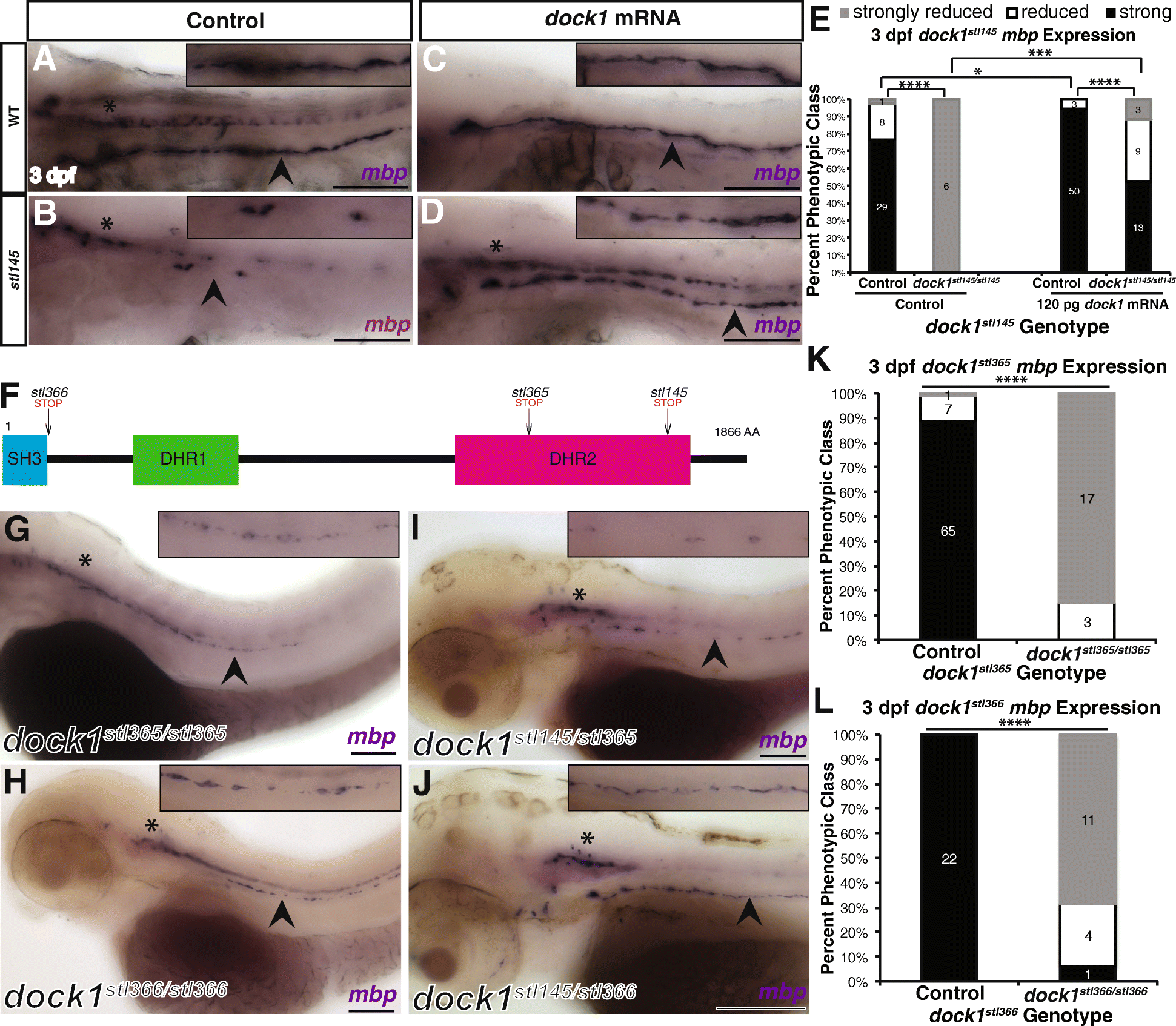
Fig. 2
Mutations in dock1 cause decreased mbp expression in the PNS. a-d) Lateral views of mbp expression by WISH at 3 dpf. Arrowheads indicate PLLn. Asterisks indicates the CNS. Inset panels show a magnified view of PLLn. Scale bars = 100 μm. a) Control larvae robustly express mbp in the PLLn (n = 29/38). b) dock1stl145 homozygous mutants exhibit strongly reduced mbp expression in the PLLn (n = 6/6). c) Control larvae injected with dock1 mRNA exhibit strong expression of mbp in the PLLn (n = 50/53). d) dock1stl145 homozygous mutants injected with dock1 mRNA robustly express mbp in the PLLn (n = 13/25). e) Quantification of the percent phenotypic classes larvae were scored for mbp expression in the PLLn at 3 dpf. Control = pooled uninjected and phenol red injected larvae. f) A schematic of the Dock1 protein with the locations of the stl366, stl365, and stl145 lesions indicated. g-j) Lateral views of mbp expression by WISH at 3 dpf. Arrowheads indicate the PLLn. Asterisks indicate the CNS. Inset panels show a magnified view of PLLn. Scale bars = 100 μm. g) dock1stl365 homozygous mutants (n = 20) and h) dock1stl366 homozygous mutants exhibit reduced mbp expression in the PLLn (n = 15/16). i) dock1stl145/stl365 compound heterozygotes and j) dock1stl145/stl366 compound heterozygotes exhibit reduced mbp expression in the PLLn. k, l) Quantification of WISH for mbp from dock1stl365 (k) and dock1stl366 (l) in-crosses based on phenotypic classes and genotypes for the respective lesions. * p < 0.05, *** p < 0.001, **** p < 0.0001, Chi-squared analysis