
|
Fig. S5
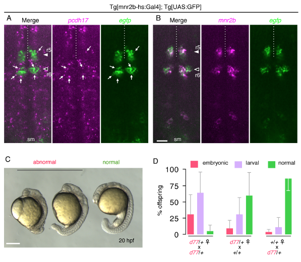
pcdh17-d77 mutation causes developmental defects. (Related to Figure 5)
(A) pcdh17 is widely expressed in the hindbrain with enhanced expression levels in specific cell populations at 44 hpf. (B) EGFP recapitulates mnr2b expression in the Tg[mnr2b-hs:Gal4]; Tg[UAS:GFP] larva. sm, spinal motor neuron. In A and B, shown are the z-stacks of three optical sections taken at the 1-μm interval (2 μm thick), which are thin enough to exclude potential colocalization of pcdh17 speckles and EGFP in different cells. (C) Examples of the embryonic defect observed at 20 hpf. (D) Incidence of developmental defects occurring during the embryonic (red) and larval (larval) stages observed among the offspring from indicated parental fish. See Supplemental experimental procedure for the criteria of phenotypic characterization. Embryonic phenotypes typically include shortened body due to defective gastrulation, which occasionally resulted in embryonic death due to yolk damage. Either of failure in forming a fully inflated swim bladder on 5 dpf or morphological abnormalities such as cardiac edema was considered as larval defects. The error bars show standard deviation. The bar indicates 50 μm in A and B, and 250 μm in C.