
|
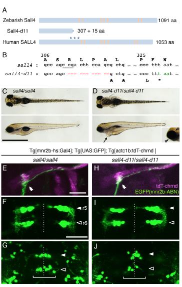
Fig. S4
sall4 knockout fish forms the neuromuscular connection between mnr2b-ABN and lateral rectus. (Related to Figure 4)
(A) The structure of zebrafish and human Sall4/SALL4 protein. The asterisks indicate the positions of mutations causing protein truncation in Okihiro/DRRS syndrome patients. The orange ovals represent the zinc finger domains. (B) The nucleotides at the position from +922 to +932, where A in the initiation codon is +1, are deleted in sall4-d11. The underlined CCG (CGG in the opposite strand) represents the protospacer adjacent motif (PAM) for Cas9 nuclease. (C, D) The dorsal (top) and lateral (bottom) views of the wild type (C) and sall4-d11 (D) larvae at 5 dpf. The arrow indicates the cardiac edema (D, bottom). Arrowheads show the eye edema (inset). (E-J) The dorsal view of the lateral rectus muscle (E, H) and the ventral (F, I) and dorsal cluster (G, J) of abducens nuclei in the wild type (left panels) and the sall4-d11 homozygote (right panels) at 5dpf. The double arrowheads indicate CEPZ. The white and black arrowheads indicate the position of r5 and r6, respectively. The brackets indicate the dorsal cluster. The dashed lines show the midline. The bars indicate 1 mm in C and D and 50 μm in others.