
|
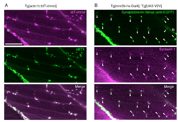
Fig. S2
Transgenic zebrafish marker lines for neuromuscular synapse. (Related to Figure 1)
(A) tdT-chrnd fluorescence represents AChR clusters in the postsynaptic endplates. The skeletal muscle of a larva at 96 hpf, treated with 1 μg/ml α-bungarotoxin Alexa FluorTM 488 conjugate (B13422, Life Technologies) for 1 hour, after a 6-hour fixation with 4% paraformaldehyde (middle, αBTX). tdT-chrnd fluorescence (top, tdT-chrnd) overlapped with αBTX signals (bottom, Merge). (B) Synaptobrevin-Venus driven by Tg[mnr2b-hs:Gal4] labels presynaptic terminals. Immunofluorescence of skeletal muscle of a larva at 96 hpf using mouse anti-EGFP (1/1000, MAB3580, CHEMICON) and rabbit anti-Syntaxin 1 (1/1000, S1172, SIGMA) antibodies. Goat anti-mouse Alexa Fluor 488 (1:1000, Molecular Probes) and goat anti-rabbit Alexa Fluor 633 (1:1000, Molecular Probes) were used as secondary antibodies. Synaptobrevin-Venus signals (top) overlapped and/or were in close apposition of presynaptic marker Syntaxin 1 (arrows). ib, intermyotomal boundary. Bar indicates 20 μm.