
|
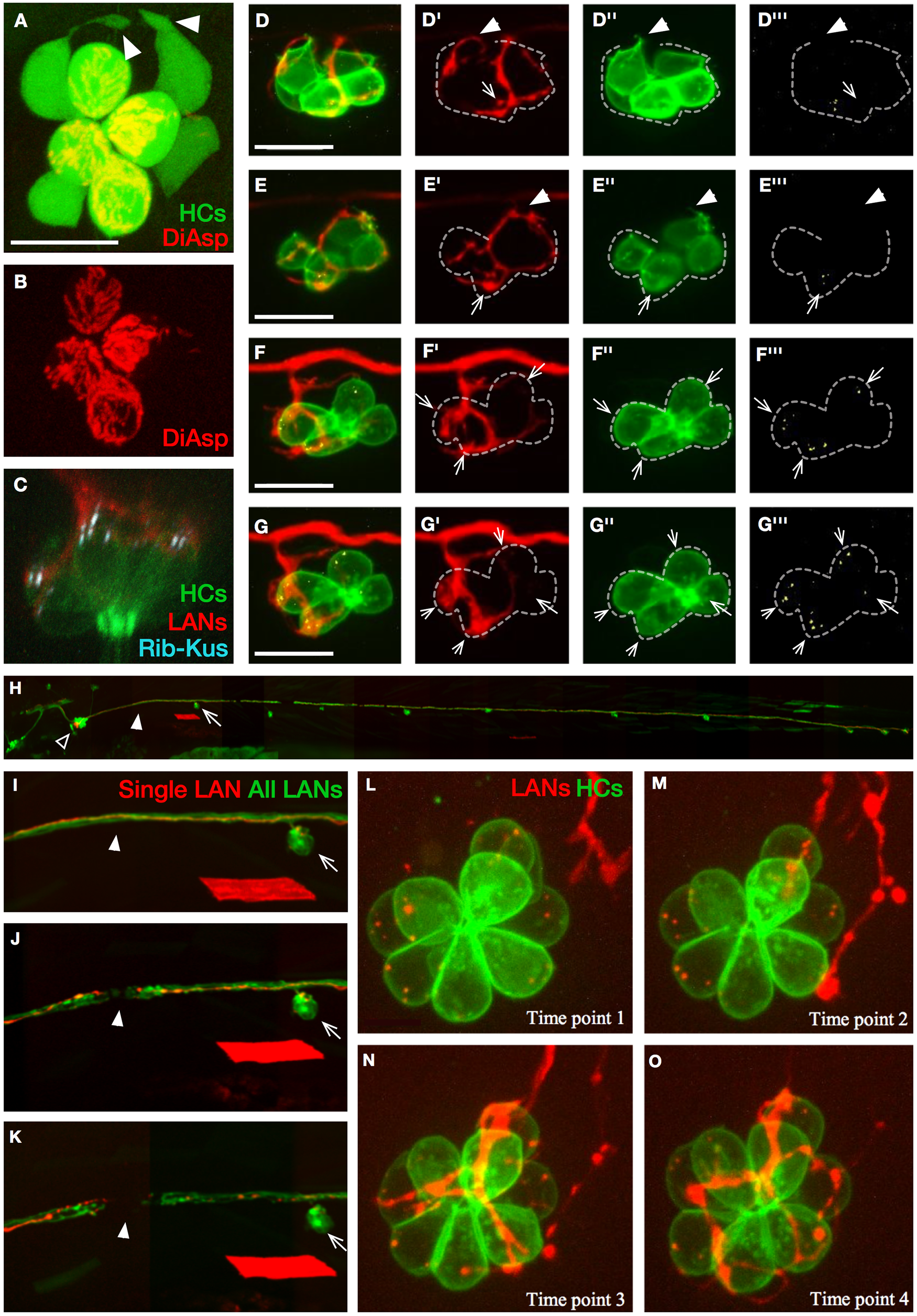
Fig. 2
Live imaging of synaptogenesis in neuromasts.
(A–B) Apical view of hair cells (green) in a horizontal neuromast of Et(krt4:EGFP)sqet4 transgenics labeled with DiAsp (red), which only penetrates into mature hair cells through mechanotransducing channels. The image reveals basal projections (white arrowheads) in 2 of the 4 immature hair cells that are DiAsp(−) (white asterisks in panel B). (C) Confocal image of a neuromast in triple-transgenics Tg[pou4f3:GAP-GFP; pou4f3:ctbp2l-mKOFP; nkhgn39d], showing juxtaposition of LAN neurites (red) with active-zone (Ribeye[+] puncta, light blue) in hair cells (green). (D–G’”) Selected time points from live imaging of innervation of hair cells (green) expressing Ribeye-Kusabira (light blue) by a singly marked LAN expressing mCherry (red), in double transgenics Tg[pou4f3:GAP-GFP; pou4f3:ctbp2l-mKOFP]. These panels were extracted from S1 Video. Ribeye(+) puncta are readily evident in hair cells (arrows) but absent from basal projections of hair cells (arrowheads in panel D–E’”). Synapses occur when LAN neurites and Ribeye(+) puncta are persistently juxtaposed over time (panel F–G’”). Dotted line outlines the hair cells. (H) Lateral view of a transgenic Tg[nkhgn39d] larval zebrafish expressing EGFP (green) in all LANs and mosaic expression of mCherry (red) in a single LAN, whose cell body (empty arrowhead) can be seen within the posterior lateralis ganglion. A neuromast (arrow) and the site of the eventual severing of the LAN peripheral axons (solid arrowheads) are indicated. The red rhomboid below the indicated neuromast is a singly marked myofiber resulting from nonspecific expression of mCherry in muscle. (I–K) Magnified views of the same fish in panel H, showing the LAN peripheral axons before laser-mediated severing (panel I), immediately after severing (panel J), and several hours after severing (panel K), evidencing the separation of the proximal and distal parts of the axon fragments. The arrows indicate the same neuromast and the solid arrowheads the site of the cut in every panel. The red rhomboid is the same myofiber in panel H. (L–O) Selected time points from live confocal imaging of regenerative innervation of mature hair cells (green) by the singly marked LAN (red). Panels were generated from S2 Video. Hair cells never produce basal projections during polarity-selective re-innervation. Scale bars are 10 μm in panel A, C, and D and 50 μm in panel I. DiAsp, 4-(4-diethy-laminostyryl)-N-methylpyridinium iodide; EGFP, enhanced green fluorescent protein; HC, hair cell; LAN, lateralis afferent neuron; Rib-Kus, Ribeye-Kusabira.