
|
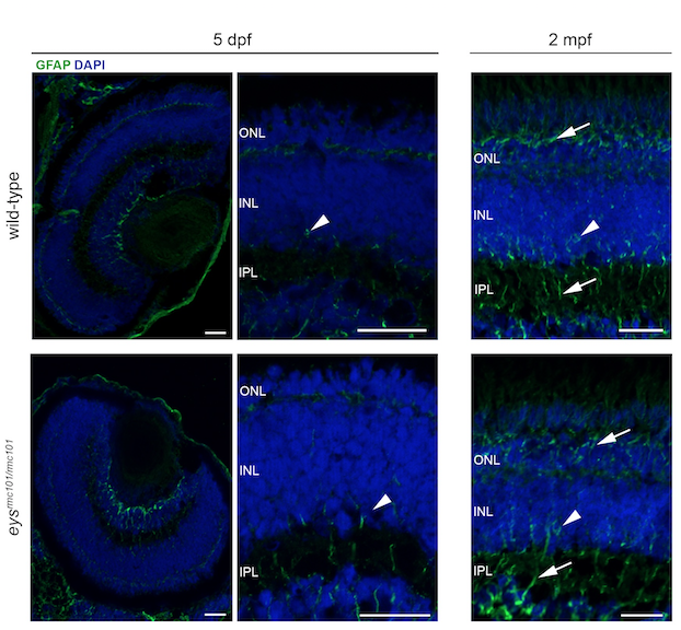
Fig. S4
Immunohistochemistry on retinal sections of wild-type and eysrmc101/rmc101 zebrafish in order to investigate Müller glia activation.
Retinal sections of wild-type and eysrmc101/rmc101 zebrafish at 5 dpf and 2 mpf stained with antibodies against GFAP (green), as a marker for Müller glia cells. Müller glia cell bodies are located in the inner nuclear layer (arrow heads) and project processes (arrows) in either direction to outer limiting membrane and inner limiting membrane. Nuclei are counterstained with DAPI (blue). INL: inner nuclear layer; IPL: inner plexiform layer; ONL: outer nuclear layer. Scale bar: 20 μm.