
Fig. 2
|
Fig. 2
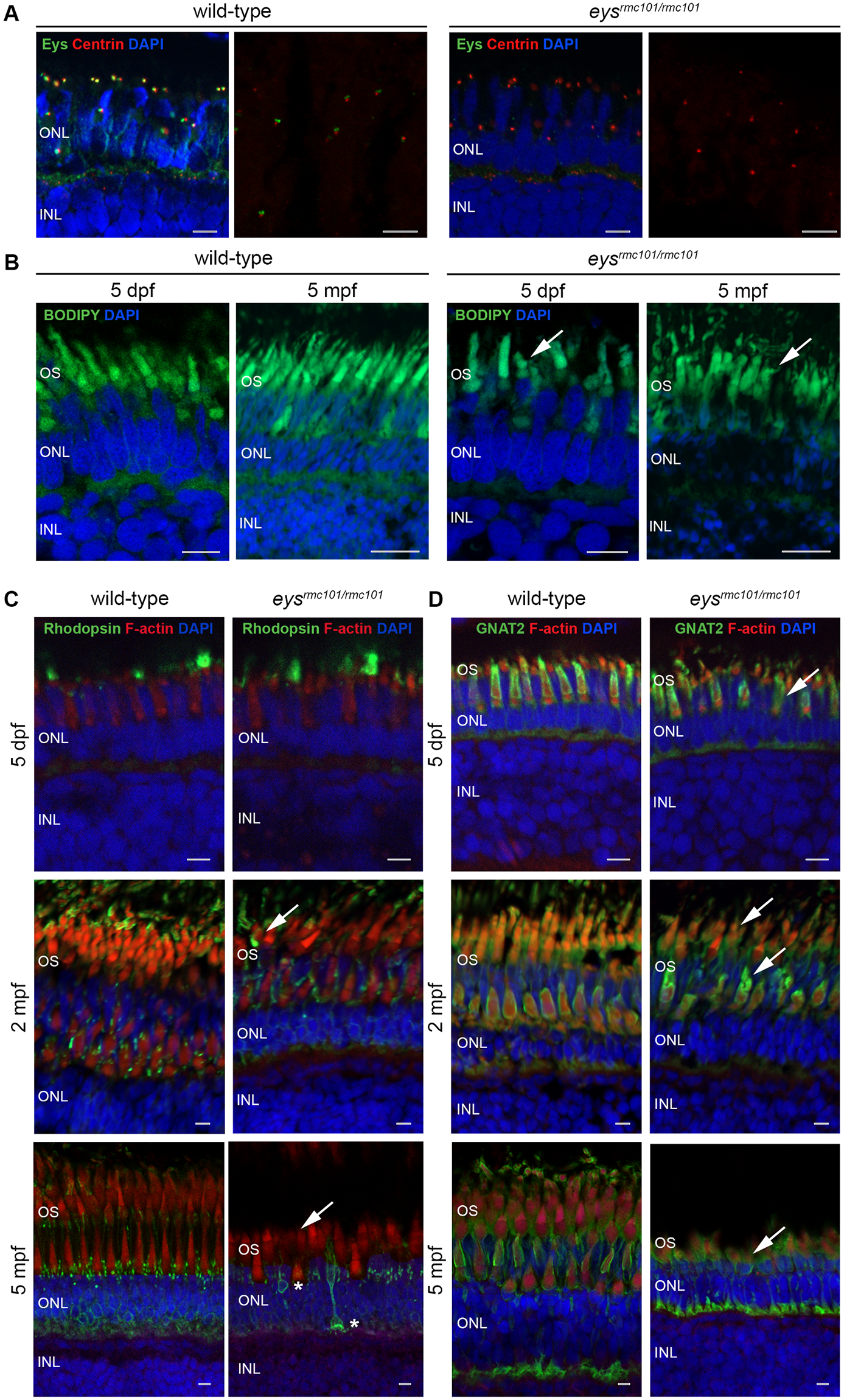
Immunohistochemistry on retinal sections of wild-type and eysrmc101/rmc101 zebrafish.
(A) Retinal sections of wild-type and eysrmc101/rmc101 zebrafish at 5 dpf and 5 mpf stained with antibodies against Eys (green) and centrin (red). (B) BODIPY (green) staining showing disorganization of photoreceptor outer segments in eysrmc101/rmc101 zebrafish (5 dpf and 5 mpf) compared to age- and strain-matched wild-type zebrafish (arrows). (C) Retinal sections of wild-type and eysrmc101/rmc101 zebrafish at 5 dpf (upper panel), 2 mpf (middle panel) and 5 mpf (lower panel) stained with antibodies against rhodopsin (green) and F-actin (red). Asterisks indicate mislocalization of rhodopsin to the inner segments and synapses of photoreceptor cells. (D) Retinal sections of wild-type and eysrmc101/rmc101 zebrafish at 5 dpf, 2 mpf and 5 mpf stained with antibodies against GNAT2 (green) and F-actin (red). Arrows indicate dysmorphic outer segments in mutant zebrafish. In all images, nuclei are counterstained with DAPI (blue). INL: inner nuclear layer; ONL: outer nuclear layer; OS: outer segments. Scale: 5 μm.