
|
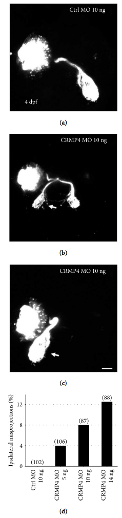
Fig. 3
Knocking down CRMP4 causes ipsilateral misprojections of retinal axons. Morpholino was injected into 1-2 cell stage embryos and retinal axons were labeled with DiI or DiD at 4 dpf. (a) A representative image demonstrating that, in control MO-treated zebrafish larvae, all retinal axons cross the midline and project into the opposite side of the tectum. (b, c) Representative images of retinal axon guidance errors in CRMP4 MO-treated larvae. A part or all of the retinal axons fail to cross the midline and misproject into the ipsilateral tectum (arrows). Note that although the axons misproject ipsilaterally, they still follow the normal optic tract and arborize into the tectum. (d) The ipsilateral misprojections of retinal axons caused by CRMP4 MOs are dose dependent. The doses of MOs are labeled under each column. The numbers in parentheses above each column indicate the amount of eyes. Scale bar: 50 μm.