
Image
|
Figure Caption
Fig. 5
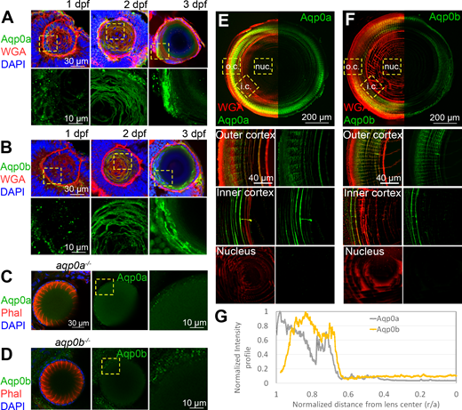
Expression of Aqp0a and Aqp0b in WT zebrafish lenses; 1 to 3 dpf eye cryosections labeled with (A) anti-Aqp0a or (B) anti-Aqp0b antibodies (green), plasma membrane label wheat germ agglutinin (WGA, red), and DAPI (blue). High power images of Aqp0a or Aqp0b labeling only were taken from regions outlined by yellow-dashed boxes. aqp0a−/− lenses labeled for F-actin (phalloidin, red) and DAPI (blue) as well as with (C) anti-Aqp0a antibody, and aqp0b−/− labeled with (D) anti-Aqp0b antibody serve as negative controls. Adult lens cryosections were labeled with (E) anti-Aqp0a or (F) anti-Aqp0b antibodies (green), and WGA (red). Equatorial lens section is shown with antibody labeling only detected in the cortex. High power images from the outer cortex (o.c.), inner cortex (i.c.), and nucleus (nuc.) are taken from regions outlined by yellow dashed boxes. (G) Normalized intensity of either Aqp0a (gray) or Aqp0b (yellow) antibody labeling in adult lenses as a function of distance from lens center. Each experiment is representative of at least six independent lenses analyzed.
Figure Data
Acknowledgments
This image is the copyrighted work of the attributed author or publisher, and
ZFIN has permission only to display this image to its users.
Additional permissions should be obtained from the applicable author or publisher of the image.
Full text @ Invest. Ophthalmol. Vis. Sci.