
|
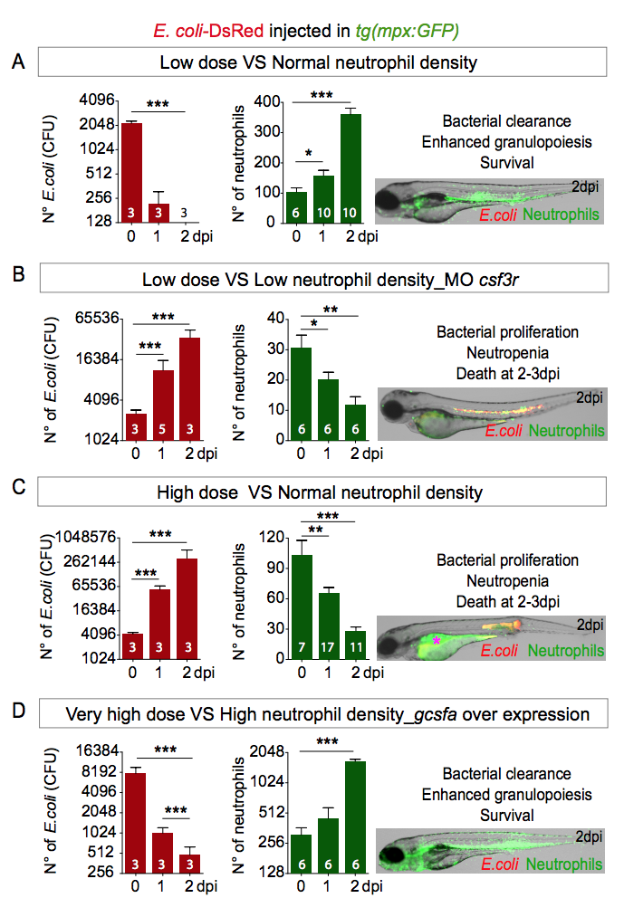
Fig. S3
Embryos need correct neutrophil density to fight notochord infection.
Two dpf tg(mpx:GFP) embryos were infected in the notochord with low dose (< 3000 CFUs) (A, B), high dose (> 4000 CFUs) (C) or very high dose (>7000 CFUs) (D). (B) To decrease neutrophil density, tg(mpx:GFP) embryos were injected at the one cell stage with the csf3r morpholino and then infected in the notochord at 2 dpf with a low dose of red fluorescent E. coli-DsRed. (D) To increase neutrophil density, tg(mpx:GFP) embryos were injected at the one cell stage with the gcsfa over-expressing plasmid and then infected in the notochord at 2 dpf with a very high dose of red fluorescent E. coli-DsRed. Charts show the quantification of CFU (red-bar charts) and of the total neutrophil number (green-bar charts) at 0, 1 and 2 dpi (Mann-Whitney test, two-tailed, Nlarvae is indicated on the columns, *p<0.05, ** p<0.01 and ***p<0.001). Larvae images are representative overlays of fluorescence (green: neutrophils and red: E. coli) and transmitted light images at 2 dpi (asterisk: auto-fluorescence of the yolk).