
|
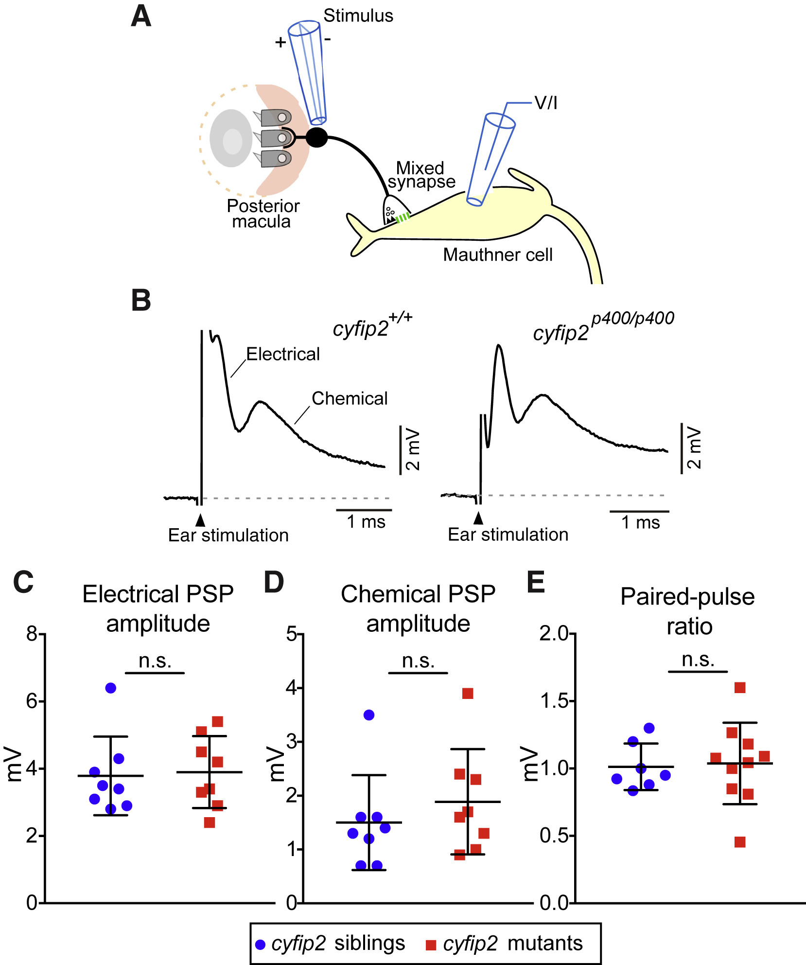
Fig. 3 VIII Nerve Excitatory Inputs to the Mauthner Cell Are Normal in cyfip2 Mutants (A) Diagram of the stimulating electrode (stimulus) adjacent to the OV posterior macula, the club-ending mixed synapse between VIII afferents and the M-cell, and the recording electrode (voltage/current [V/I]) on the M-cell. (B) Representative traces of M-cell synaptic responses after stimulation of VIII afferents in cyfip2+/+ (left) and cyfip2p400/p400 (right) larvae at 5 dpf. The stimulation artifact has been truncated for clarity, and the electrical and chemical components are indicated. (C and D) Mean amplitude of M-cell electrical (C) and chemical synaptic responses (D) ± SD (n = 8 siblings, 8 mutants; p = 0.78, 0.29, Mann-Whitney test). (E) Paired-pulse ratios were unaltered in cyfip2p400/p400 larvae (p = 0.76, Mann-Whitney test).