
|
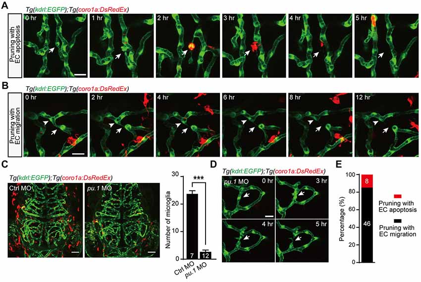
Fig. 3
Role of microglia in EC apoptosis-accompanied brain vessel pruning. (A) In vivo time-lapse confocal images showing that, during EC apoptosis-accompanied vessel pruning, a microglial cell (red) migrated to, engulfed and cleaned the apoptotic EC (white arrow). The Tg(coro1a:DsRedEx);Tg(kdrl:EGFP) larvae at 3–3.5 dpf were used. (B) In vivo time-lapse confocal images showing that, during EC migration-accompanied vessel pruning (white arrow), there was no obvious interaction between microglia (red) and the migrating EC (white arrowhead). (C) Representative projected confocal images (left) and summary data (right) showing that knockdown of pu.1 significantly diminished the number of microglia (red) in the brain at 3 dpf. (D) In vivo time-lapse confocal images showing that EC apoptosis-accompanied brain vessel pruning (arrow) still occurred in pu.1 morphants. (E) Summary of the percentages of EC apoptosis- and migration-accompanied brain vessel pruning from pu.1 MO-injected Tg(coro1a:DsRedEx);Tg(kdrl:EGFP) (n = 30 larvae). The numbers on the bars represent the number of animals (C) or pruned vessels (E) examined. Data are shown as mean ± SEM. ***p < 0.001 (two-tailed unpaired Student’s t-test). Scales: 20 μm (A,B), 50 μm (C) and 15 μm (D).