
|
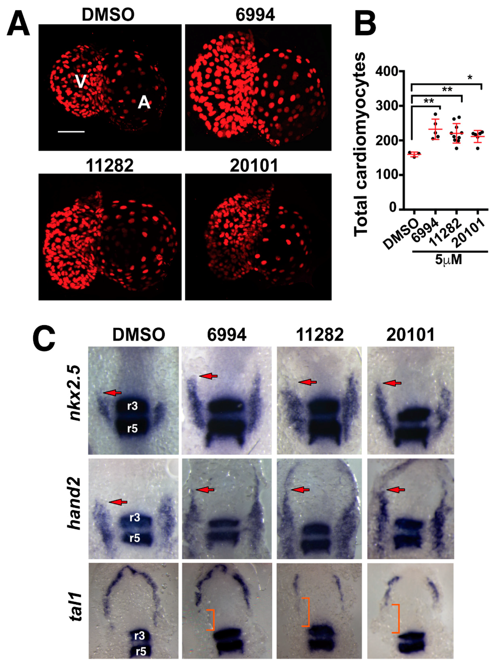
Fig. 4
Fgf hyperactivators regulate heart size by increasing cardiac progenitor populations. (A) Fluorescence micrographs of cardiomyocyte nuclei from 72 hpf transgenic larvae treated for somite stages one through eight with 5 µM BCI, ST006994, ST011282, or ST020101. Images are representative for each compound. (B) The quantification of cardiomyocytes after small molecule treatment. All three compounds significantly increased the numbers of cardiomyocytes in the developing zebrafish heart. Each point represents a single larvae. (C) Whole mount in situ hybridization shows the expression of cardiac progenitor markers (nkx2.5 and hand2) and endothelial progenitors (tal1) after compound treatment. egr2b was used to mark the hindbrain rhombomeres 3 and 5 (r3 and r5). Arrows mark the rostral most domain of the cardiac progenitor population. Brackets mark the region of decreased endothelial tal1 expression.