
|
Fig. S4
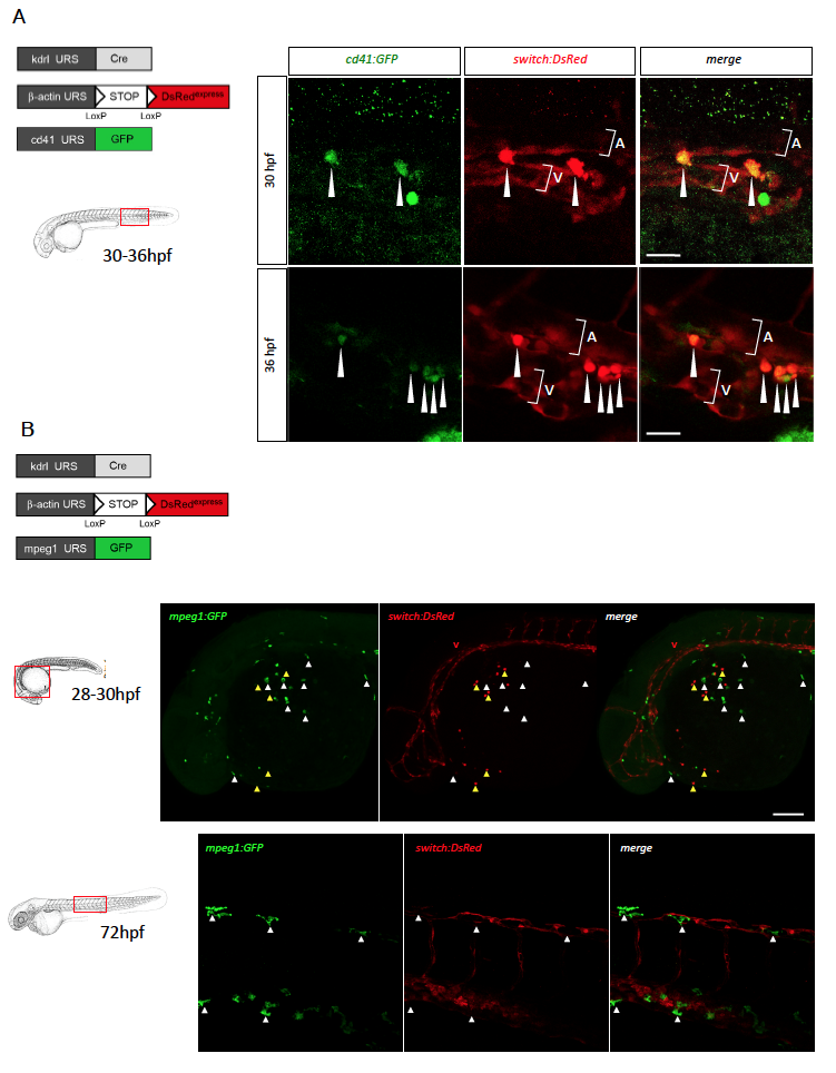
Zebrafish EMPs, but not primitive macrophages, derive from an endothelial precursor, related to Figure 3
(A) Triple transgenic kdrl:Cre;bactin2:loxP-STOP-loxP-DsRedexpress;cd41:GFP embryos were analyzed by confocal imaging at 30hpf (upper panels) and 36hpf (lower panels). Shown are GFP, DsRed and merged images of posterior blood island, posterior to the yolk tube extension. In this area, cd41+ erythro-myeloid precursors express both GFP and DsRed (white arrowheads). A= aorta, V= vein. Scale bar = 25μm.
(B) Immunostaining and confocal microscopy analyses of triple transgenic Tg(kdrl:Cre;bactin2:loxP-STOPloxP- DsRedexpress;mpeg1:GFP) embryos at 28-30hpf (upper panels). Shown are GFP, DsRed and merged images of the rostral region, demonstrating the absence of co-localization between GFP+ pMFs emerging on the yolk (white arrowheads) and the DsRed reporter transgene. In contrast, DsRed expression is observed in the vasculature (V) and in hematopoietic cells subsequently identified as primitive neutrophils (yellow arrowheads). Scale bar = 100μm. Similar analyses performed at 72hpf (lower panels), showing GFP+DsRedpMFs spreading over embryonic tissues, here, in the trunk region. Scale bar = 50μm.