
Image
|
Figure Caption
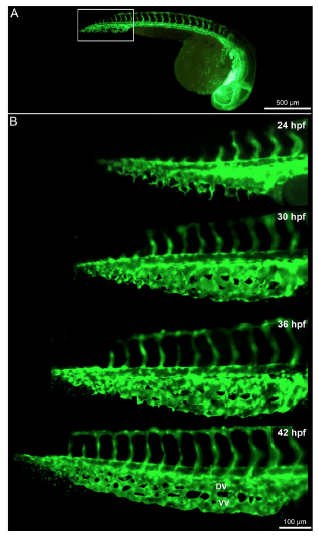
Fig. 1
Time lapse in vivo images of the caudal vein plexus (CVP) from 24-42 hpf. (A) Overview of the blood vessels in Tg(fli1a:eGFP)y7 of the whole zebrafish embryo with the highlighted region (white box) showing the developing CVP. (B) In vivo still images of the CVP from selected ages between 24 hpf and 42 hpf indicating the mode of angiogenesis. Sprouting and anastomoses are evident at 24 hpf while pillar formation starts from 30 hpf increasing in intensity towards 42 hpf.
Acknowledgments
This image is the copyrighted work of the attributed author or publisher, and
ZFIN has permission only to display this image to its users.
Additional permissions should be obtained from the applicable author or publisher of the image.
Full text @ Sci. Rep.