
Fig. 5
- ID
- ZDB-IMAGE-180823-9
- Genes
- Antibodies
- Publication
- Moreno et al., 2018 - Investigation of Islet2a function in zebrafish embryos: Mutants and morphants differ in morphologic phenotypes and gene expression
- All Figures
- Figures for Moreno et al., 2018
|
Fig. 5
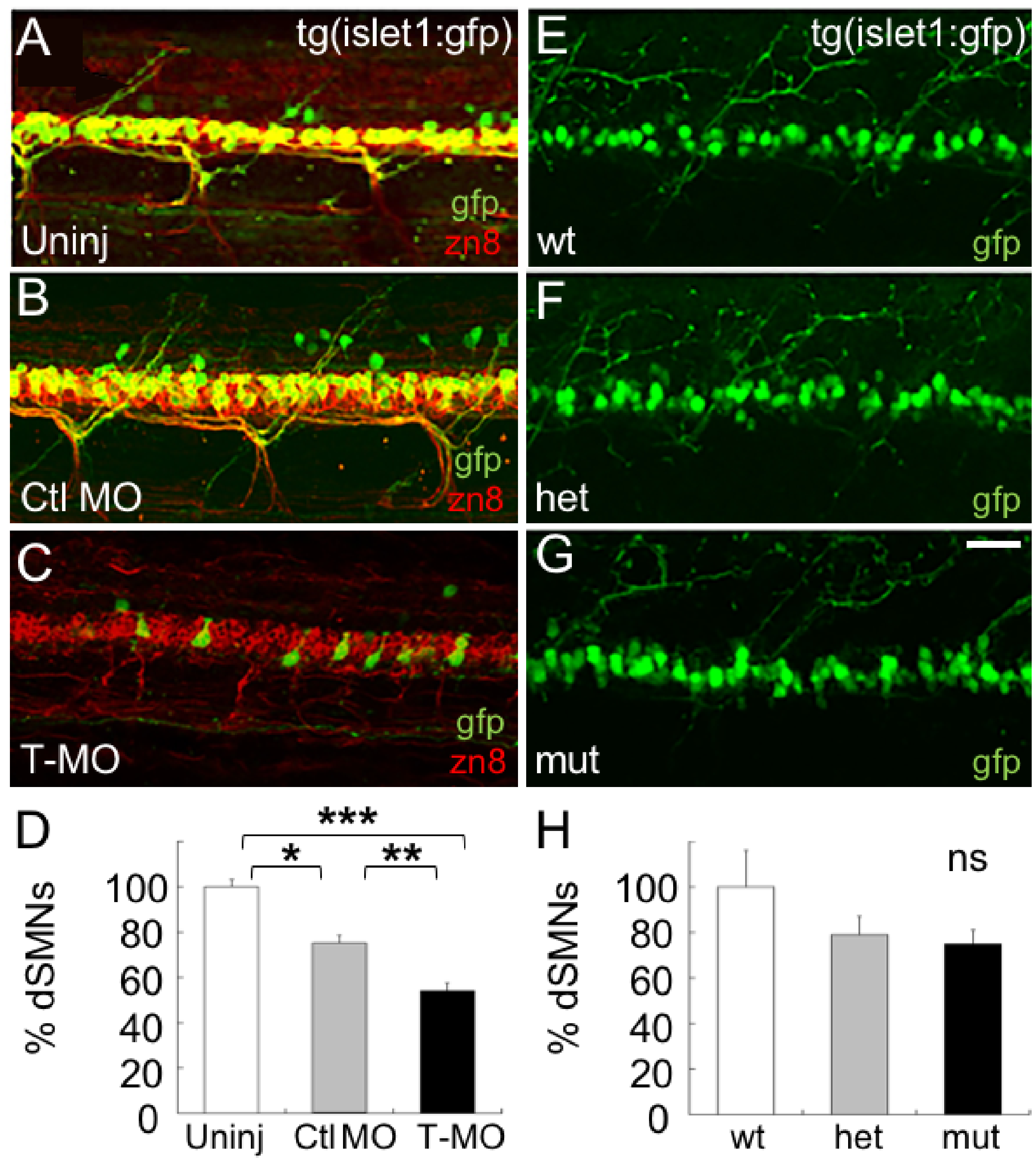
Islet2a morphant, but not mutant, larvae have a reduced number of dSMNs.
(A-C) In 72 hpf tg(islet1:gfp) larvae, dSMNs expressed gfp in their somas and axons. In addition, the zn8 antibody recognized the neurolin protein (red), expressed on SMN somas and axons [38]. (A) In uninjected 72 hpf larvae, the majority of zn8+ neurons also expressed gfp. (B) Following injection of the control MO (Ctl MO), zn8+ (red) neurons continued to express gfp. In addition, dSMNs developed normally with respect to axon morphology (arrowhead), as assessed by zn8 immunolabeling (red). (C) Injection of the T-MO led to a decrease in the number of zn8+ neurons that coexpressed gfp. Scale bar in A, for A-C: 50 μm. (D) In tg(islet1:gfp) 72 hpf larvae, the numbers of gfp+ somas were reduced by injection of the Ctl MO (n = 17) and further reduced by injection of the T-MO (n = 20) compared to uninjected embryos (n = 10). ***, p<0.0001; **, p<0.0003; 8, p<0.0006; ANOVA with post-hoc Bonferroni. (E-G) In live 72 hpf tg(islet1:gfp) larvae, dSMNs appeared normal in number and morphology in homozygous mutant (mut; G) compared to heterozygous (het; F) and homozygous wildtype (wt; E) 72 hpf embryos. (H) Cell counts indicated that the number of dSMN somas was not reduced in mutant (n = 9) compared to wildtype (n = 5) or heterozygous (n = 8) islet2a 72 hpf embryos.