
|
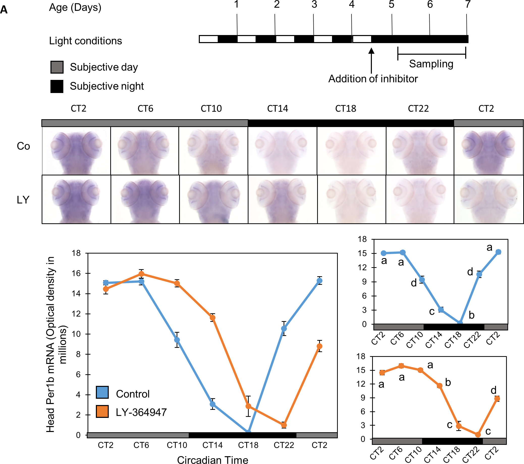
Fig. 5
Per1b mRNA circadian expression pattern in zebrafish larvae is phase-shifted by TGF-β inhibition.
Zebrafish larvae were treated with TGF-β inhibitor LY-364947 (20 μM), and the expression pattern of Per1b was evaluated by whole mount ISH. Per1b expression was detected throughout the head region and its circadian expression pattern was altered in the presence of the TGF-β inhibitor, exhibiting a phase delay of circadian expression in comparison to a control group (DMSO). Per1b mRNA expression was significantly affected by sampling time (p<0.001, two-way ANOVA), and by an interaction between treatment and sampling time (p<0.001, two-way ANOVA) (n = 15/group). (A) Schematic representation of the experimental design. The horizontal bars represent the light conditions before and during sampling; white boxes represent light and black boxes represent dark periods. Bottom panel: whole mount ISH signals for Per1b mRNA (dorsal views of the heads) of representative specimens. Grey bars represent subjective day and black bars represent subjective night. Circadian times are indicated for each sample. CT0 corresponds to “subjective lights on”, CT12 to “subjective lights-off”. (B) Left: Quantification of signal intensities in the heads of treated and control larvae. Values represent the mean ± SE optical densities of the head signals. Right: Different letters represent statistically different values within each treatment (p<0.05, one-way ANOVA, Tukey’s test). This experiment was repeated twice, resulting in similar outcomes. The represented results are of one experiment.