
|
Fig. 1
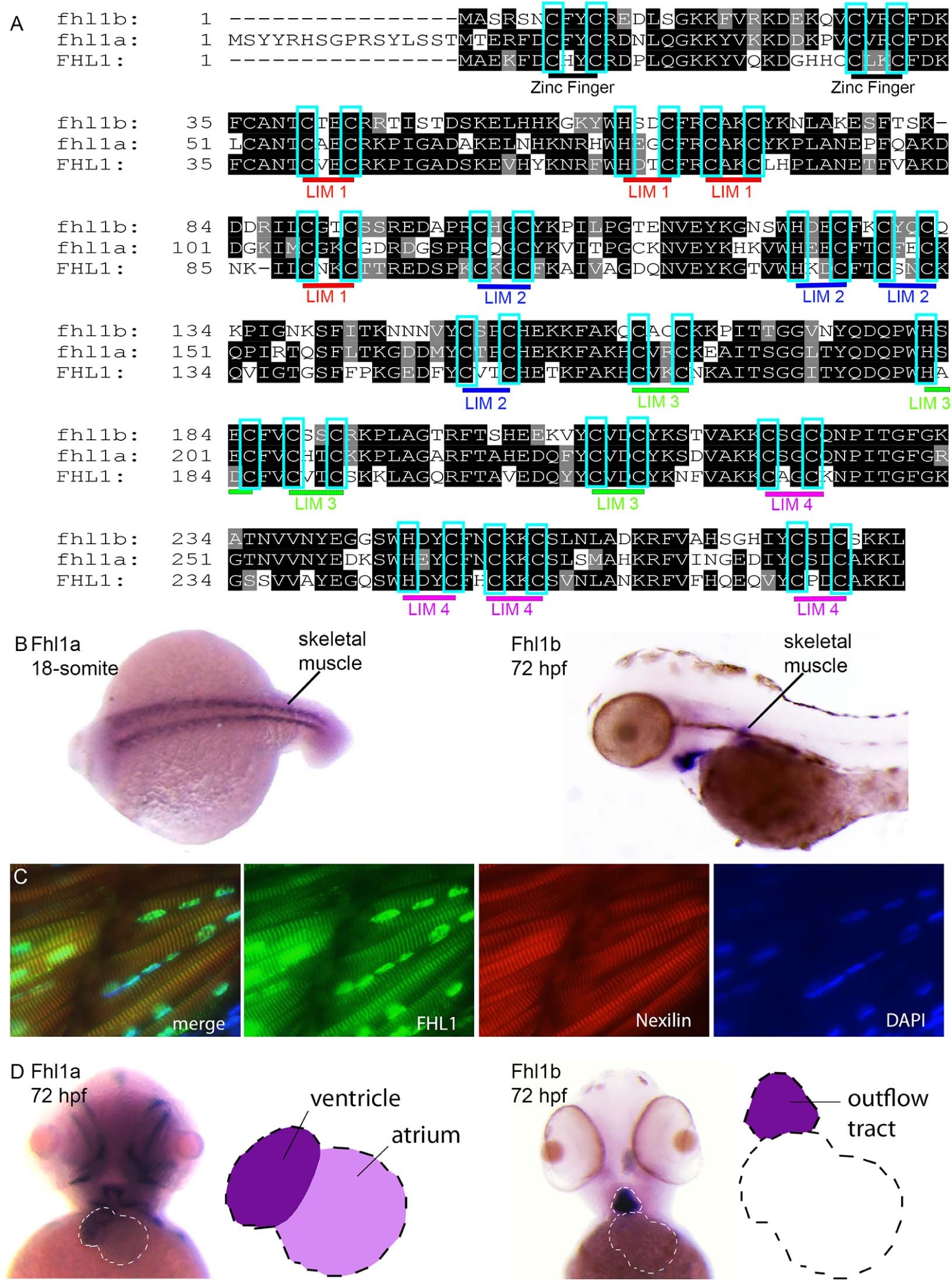
(A) Box shade diagram of amino acid sequence of human FHL1 and both zebrafish orthologs fhl1a and Fhl1b. Amino acid sequences of zebrafish Fhl1a and Fhl1b are highly conserved from zebrafish to humans, showing a 67% for Fhl1a and 60% for Fhl1b overall amino acid identity with human FHL1, and a 100% conservation within the essential Zinc-binding LIM domains (light blue boxes). (B) Whole-mount RNA in situ hybridization using a fhl1a (left) and fhl1b (right) antisense probe. Both, fhl1a and fhl1b, localize to skeletal muscle. fhl1a is detectable abundantly already at the 18-somite stage of embryonic development in the skeletal muscle, whereas fhl1b localizes to slow muscle cells and the pectoral fin muscles at 72 hpf. (C) Immunhistochemistry of zebrafish skeletal muscle. Merge of FHL1 and Nexilin immunohistochemistry and DAPI staining. FHL1 localizes to z-discs and nucleus. Nexilin localizes to z-discs. DAPI staining of the nucleus. (D) Whole-mount RNA in situ hybridization using a fhl1a (left) and fhl1b (right) antisense probe. Both, fhl1a and fhl1b, localize to the heart.