
Image
|
Figure Caption
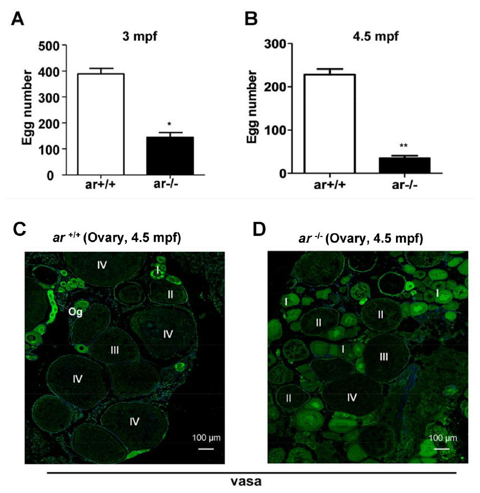
Fig. S4 (A, B) At 3 or 4.5 mpf, ovary eggs were fewer in ar -/- females compared to wildtype siblings (ar +/+) (n=5 groups, which were measured everyday for 2 weeks). (C, D) Immunofluorescent staining using anti-vasa antibody identified different stages of oogenesis in ovaries from wildtype (ar +/+) and homozygous (ar -/-) (ar ihb1225/ihb1225) zebrafish at 4.5 mpf. I, stage I oocyte; II, stage II; III, stage III; IV, stage IV. Mpf, months post fertilization.
Figure Data
Acknowledgments
This image is the copyrighted work of the attributed author or publisher, and
ZFIN has permission only to display this image to its users.
Additional permissions should be obtained from the applicable author or publisher of the image.
Full text @ Oncotarget