
|
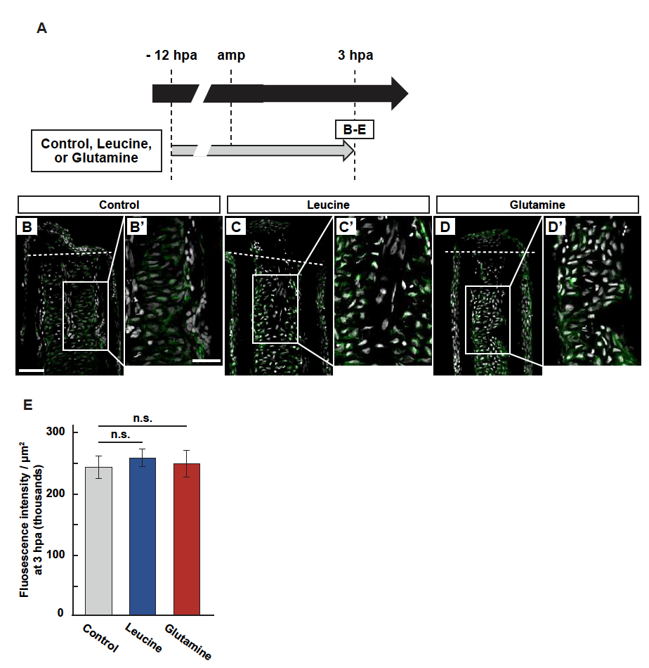
Fig. S8
Leucine or glutamine treatment has no effect on S6K activation.
(A) Experimental scheme of leucine or glutamine treatment from -12 to 3 hpa. (B-E) Longitudinal ray sections and quantification of p-S6K fluorescence intensities per area that consists of the whole regenerates and 500 µm below the amputation plane in control, leucine-, or glutamine-treated fin regenerates at 3 hpa; p-S6K and nuclei were visualized by immunohistochemical staining and DAPI staining, respectively (n = 5). Representative images (B-D’) used for quantification are shown in E. White dashed lines indicate the amputation planes. Scale bars: 50µm. n.s.: not significant. Error bars represent the standard error.