
Image
|
Figure Caption
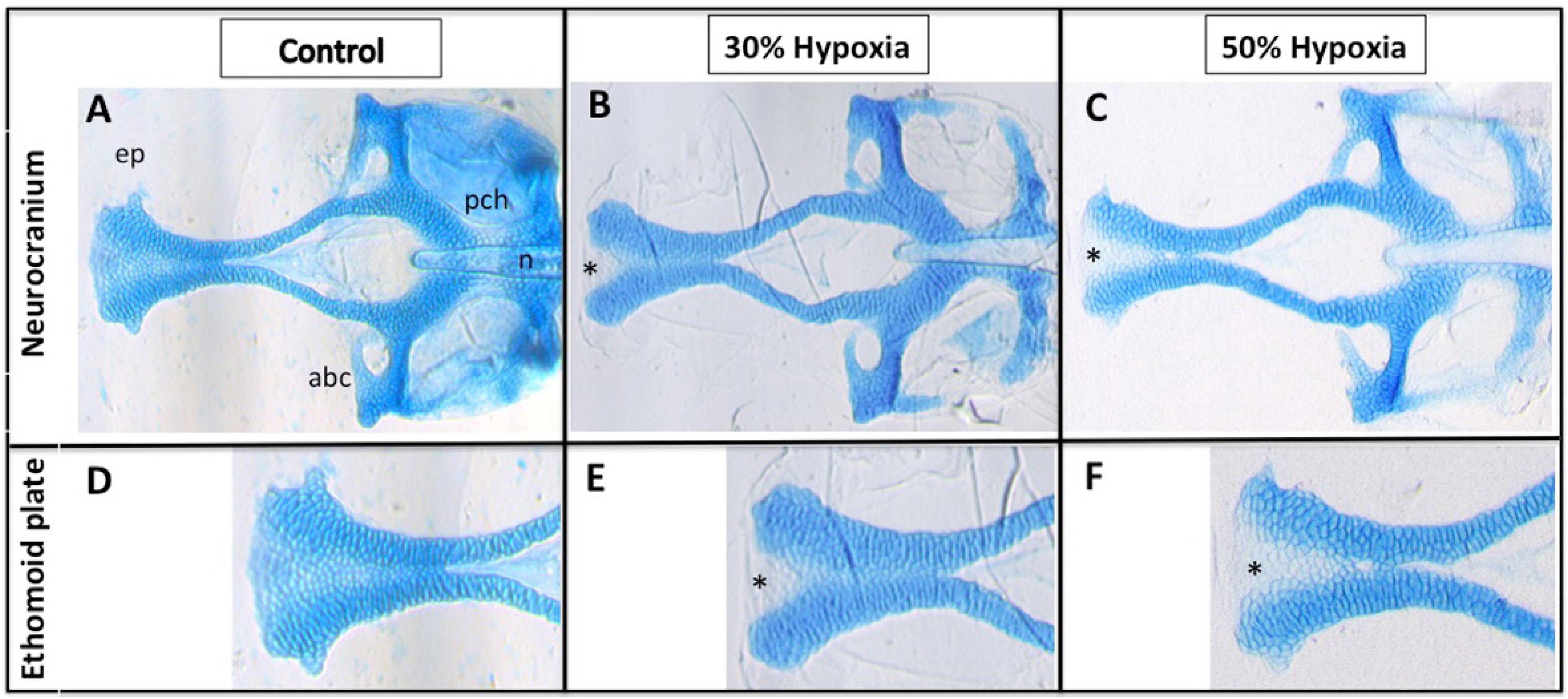
Fig. 2
Hypoxia results in craniofacial defects. (A-F) Ventral view of 5 dpf larvae stained with Alcian Blue; (A-C) Dissection of the neurocranium; (D-F) Closer view of the ethmoid plate; (B, C, E and F) Morphological alterations in the anterior area of the ethmoid plate, including a gap in the anterior edge forming a cleft; (C and F) Results of a slightly more severe phenotype in the ethmoid plate (deeper cleft). abc= anterior basicapsular commissure; ep= ethmoid plate; n= notochord; pch= parachordal; * indicates cleft in the ethmoid plate
Figure Data
Acknowledgments
This image is the copyrighted work of the attributed author or publisher, and
ZFIN has permission only to display this image to its users.
Additional permissions should be obtained from the applicable author or publisher of the image.
Full text @ J Appl Oral Sci