
|
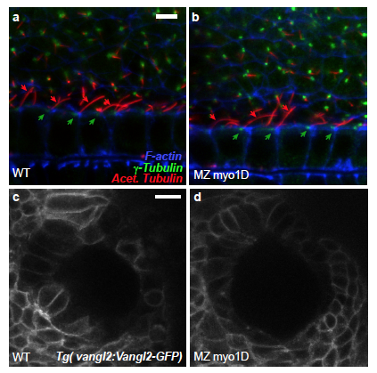
Fig. S10
Myo1D is not required for floor plate cilia orientation and Vangl2 localisation a,b, In the floor plate of 22 somite stage WT controls (a), 85.3% of the analyzed basal bodies (n=116) are posteriorly localized (green arrows) and 90.6% of the cilia posteriorly tilted (n=117, red arrows). Similarly, 80.5% of the basal bodies (n=164) and 96.8% of the cilia (n=157) are oriented towards the posterior in MZ myo1D mutants (b). Data were obtained from 18 WT and 22 MZ myo1D mutant embryos. Lateral views of the floor plate, anterior to the left. c,d, In the Left-Right organizer of both WT (n=15) and MZ myo1D mutant (n=20) embryos, Vangl2-GFP displays a similar localisation at the cell cortex. Dorsal views of 8 somites stage KVs, anterior up. Scale bars: 5 μm in a,b. 15 μm in c,d.