
Image
|
Figure Caption
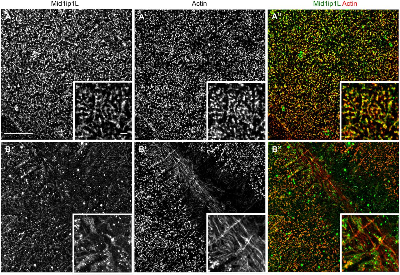
Fig. 9
Mid1ip1L protein localizes with cortex and furrow actin. (A-B″) SIM images of single and merged channels showing partial colocalization of Mid1ip1L protein with F-actin in wild-type embryos. (A-A″) At the cortex, Mid1ip1L protein partially colocalizes with the cortical F-actin. (B-B″) At the furrow, Mid1ip1L protein also partially colocalizes with transverse F-actin at furrow distal ends, which corresponds to furrow F-actin indentations (see also Fig. S5). Insets show a magnified view within the field. Scale bar: 10 μm in A-B″; 1 μm in insets.
Acknowledgments
This image is the copyrighted work of the attributed author or publisher, and
ZFIN has permission only to display this image to its users.
Additional permissions should be obtained from the applicable author or publisher of the image.
Full text @ Development