
|
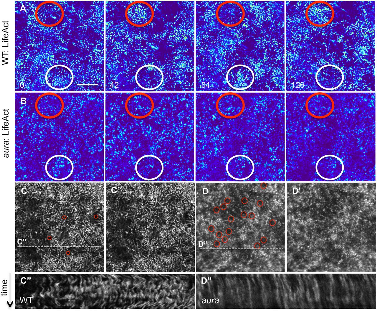
Fig. 6
Dynamic waves of cortical F-actin depend on aura/mid1ip1 function. (A,B) In live wild-type embryos, cortical F-actin shows cyclical patterns of apparent enrichment, whereas this network appears static in aura mutants (B) (in each case, red and white circles highlight two different cortical locations). Still images of LifeAct transgenic embryos spaced 42 s apart (Movie 1). (C-D′) The F-actin network towards the end of the movie sequence highlights that F-actin forms a punctate pattern as well as numerous round F-actin structures (red circles) in mutants (D,D′) that are present in lower numbers in wild type (D,D′). (C″,D″) Kymographs of cortical F-actin (data from dashed lines in C′,D′, representing 70 time points in a 140 s period) show lateral undulations in wild type and a static network in aura mutants. Scale bar: 10 μm.