
|
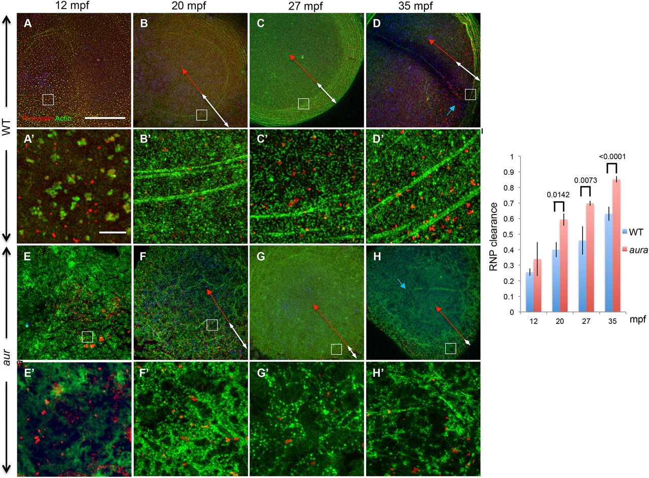
Fig. 4
Increased outward movement of germ plasm RNPs in aura/mid1ip1l mutants. (A-D) In wild-type embryos, RNPs, which are initially distributed throughout the blastodisc, progressively accumulate as a peripheral band coincident with F-actin arc formation and peripheral movement. (E-H) In aura mutants, F-actin arcs fail to form and the field of RNPs narrows at a faster rate. White double-headed arrows highlight the RNP field; red double-headed arrows indicate the distance from the center of the embryo to the inner rim of aggregates at the RNP field. Blue arrows (D,H) indicate furrow F-actin. A′-H′ are magnified regions of A-H at the location indicated by insets. Scale bars: in A, 100 μm for A-H; in A′, 10 μm for A′-H′. (I) Quantitation of outward RNP movement, with the location of the RNP aggregating front determined upon magnification of the image. Significance was determined using an unpaired t-test. Data are mean±s.e.m.