
Image
|
Figure Caption
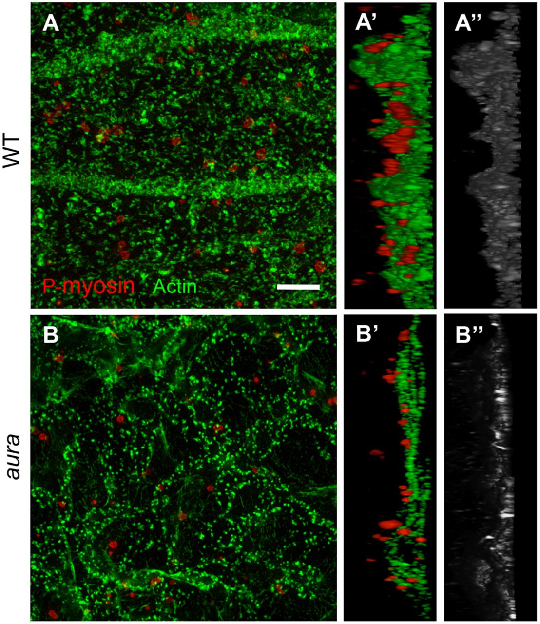
Fig. 3
Germ plasm RNP aggregates associate with Mid1ip1L-dependent cortical F-actin contractions. (A-B″) SIM images of cortical F-actin and germ plasm RNPs (anti-P-myosin) in wild type (A-A″) and aura mutants (B-B″), with orthogonal views (A′,B′, F-actin/RNPs; A″,B″, F-actin alone). Scale bar: 5 μm.
Figure Data
Acknowledgments
This image is the copyrighted work of the attributed author or publisher, and
ZFIN has permission only to display this image to its users.
Additional permissions should be obtained from the applicable author or publisher of the image.
Full text @ Development