
|
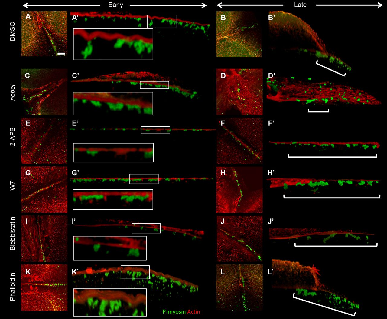
Fig. 9
Furrow F-actin contractions associated with germ plasm RNPs depend on calcium, Calmodulin and myosin. (A-L) z-projections from early and late furrows (second and first furrows at 60 mpf, respectively). (A′-L′) Lateral views showing F-actin contractions and RNPs. Control treated embryos exhibit F-actin indentations (A,A′, 5/7) and subsequent RNP distal enrichment (B,B′, 4/5). nebel mutants and 2-APB-treated wild-type embryos exhibit reduced F-actin contractions (C,C′, 2/10) and failed distal RNP enrichment (D,D′, 3/10). (E-J) Various inhibitors result in reduced F-actin indentations and RNP distal enrichment: IP3 receptor inhibitor 2-APB (E-F′, 1/5, 0/4); Calmodulin inhibitor W7 (G-H′, 0/4, 0/4); myosin inhibitor blebbistatin (I-J′, 2/5, 2/7). Phalloidin-treated embryos exhibit F-actin indentations (K,K′, 5/8) but late furrows exhibit incomplete RNP distal enrichment (L,L′, 0/14). Insets (A′,C′,E′,G′,I′,K′) are magnifications of the boxed regions. Brackets (B′,D′,F′,H′,J′,L′) highlight regions of RNP distribution. Scale bar: 10 μm in A for A-L.