
|
Fig. 3
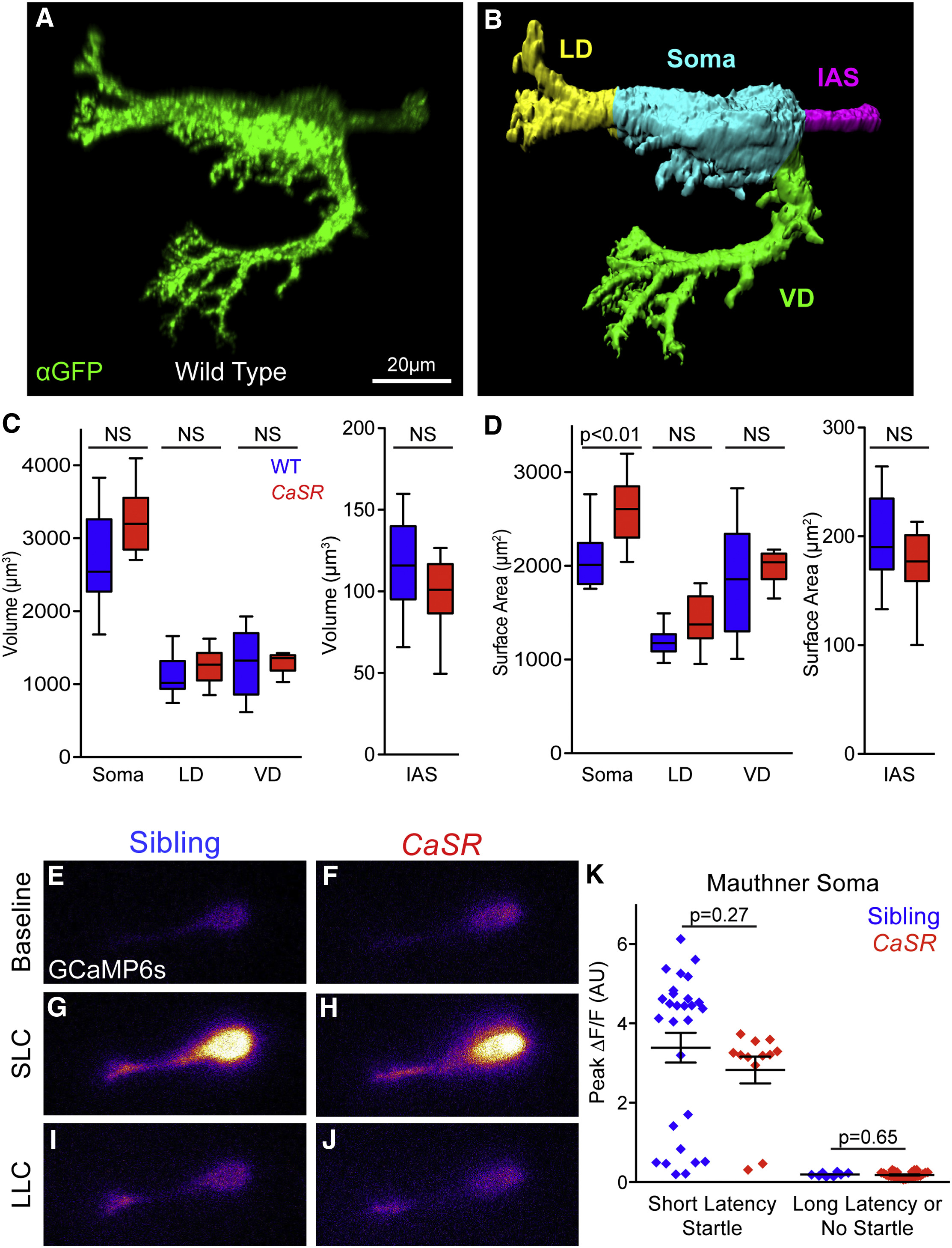
Mauthner Morphology and Function Are Largely Unperturbed in CaSR Mutants
(A) Representative projection of wild-type Mauthner-neuron-expressing membrane-targeted gap43-citrine, stained with anti-GFP. Non-Mauthner labeling was thresholded, and neurons were reoriented for clarity.
(B) Surface of wild-type Mauthner neuron from (A) segmented into the lateral dendrite (LD, yellow), ventral dendrite (VD, green), initial axon segment (IAS, magenta), and soma (cyan) for morphological quantification.
(C and D) Quantification of the volume (C) and surface area (D) of segmented Mauthner neuron regions (n = 15 sibling and 12 CaSR mutant neurons).
(E–J) Representative GCaMP6s fluorescence in Mauthner neurons of sibling (E, G, and I) and CaSR mutant (F, H, and J) larvae. Baseline fluorescence immediately prior to stimuli (E and F), and peak fluorescence during SLC (G and H) or LLC (I and J).
(K) Peak ΔF/F in Mauthner soma during responses of sibling (blue) and CaSR mutant (red) larvae to 13-dB acoustic stimuli. n = 28 sibling SLC responses, 12 CaSR SLC responses, 8 sibling LLC responses, and 26 CaSR LLC responses.
Error bars indicate SEM. See also Figure S5 and Table S2.