
Image
|
Figure Caption
Fig. 5-S2
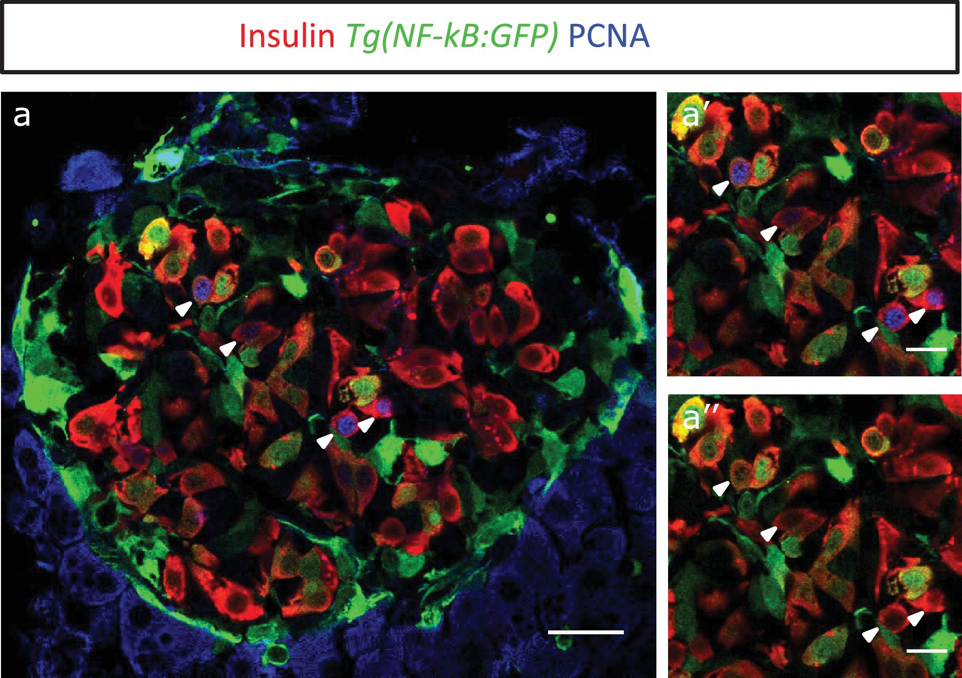
Proliferating cell nuclear antigen (PCNA) antibody staining shows that NF-kB:GF Phighbeta-cells proliferate less than NF-kB:GFP low beta-cells.
(a) Islets were stained for PCNA to mark the proliferating beta-cells in Tg(NF-kB:GFP) animals at 3 mpf. The confocal image (single plane) shows an overview of a section through the islet. The sections were stained with an insulin antibody (red), a GFP antibody (green) and PCNA (blue). (a’–a’’) The insets show higher magnification images. PCNA can be observed in some of the GFPlow cells (white arrow-heads).
Acknowledgments
This image is the copyrighted work of the attributed author or publisher, and
ZFIN has permission only to display this image to its users.
Additional permissions should be obtained from the applicable author or publisher of the image.
Full text @ Elife