
|
Fig. S5
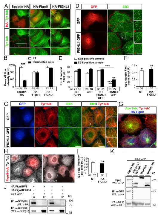
Fignl1 overexpression displaces +TIPs from MT plus ends through its specific interaction with EB1/3. (A) Overexpression of HA-tagged zebrafish spastin (spastin-HA), zebrafish Fignl1 (HA-Fignl1), and human fignl1 (HA-FIG NL1) in COS-7 cells and immunolabeling with HA (red) and tyrosinated tubulin (green) antibodies 24 hpt. Transfected cells are delineated by red dashed lines. Bottom panels represent higher magnifications of boxed regions in corresponding panels. (B) Mean MT fluorescence intensity related to cell area. (C and D) Overexpression of GFP-tagged human FIG NL1 (FIG NL1-GFP) or GFP alone (GFP) in COS-7 cells followed by immunolabeling by using GFP (C, blue), EB1 (C, green), and tyrosinated tubulin (C, red) antibodies or GFP (D, red) and EB3 (D, green) antibodies at 24 hpt. Transfected cells are delineated by blue (C) or red (D) dotted lines. Right panels represent higher magnifications of the indicated boxed region on adjacent left panels. (E) Mean number of EB1- and EB3-positive comets per 1,000 μm2. (F and G) COS-7 cells transfected with HA-Fignl1 and immunolabeled with HA, acetylated (Ace Tubulin), and tyrosinated tubulin (Tyr tubulin) antibodies. (F) Mean acetylated tubulin fluorescence intensity related to cell area. (G) Transfected cells are delineated by blue dashed lines. (H) COS-7 cells transfected with HA-Fignl1 were treated 24 hpt with 20 μM nocodazole for 15 min or with DMSO as a control and stained with HA and tyrosinated tubulin antibodies. Bars: (A, C and D [main images], F, and H) 20 μm; (C and D, insets) 10 μm. (I) Mean MT fluorescence intensity related to cell area. NT, nontransfected. The total number of cells (n) analyzed per condition in three independent experiments is mentioned under the corresponding histogram bar. **, P ≤ 0.01; ***, P ≤ 0.001; unpaired two-tailed t test. Error bars are SEM. (J) Co-IP of HA-Fignl1 or mutated HA-Fignl1E488A with EB1-GFP. COS-7 cells cotransfected with EB1-GFP and HA-Fignl1 or HA-Fignl1E488A were lysed in RIPA buffer 24 hpt. Co-IP assays were performed with a GFP antibody, and immunoprecipitated (IP) proteins were revealed by WB using HA or GFP antibodies. WT and ATP hydrolysis-deficient zebrafish Fignl1 equally coimmunoprecipitate with the core +TIP EB1. (K) co-IP of HA-tagged Fignl1 isoforms and their homologues Fign and spastin with EB3-GFP. COS-7 cells were cotransfected with EB3-GFP and FL-Fignl1–HA, Fignl1Δ1–113-HA, HA-Fign, or spastin-HA. Co-IP assays were performed 24 hpt with a GFP antibody, and immunoprecipitated proteins were revealed using either HA or GFP antibody. m: mouse; Rb, rabbit.