
|
Fig. S4
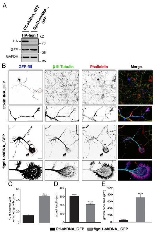
Knockdown of fignl1 in cultured mammalian neurons affects GC morphology and MT organization as in zebrafish neurons. (A) Knockdown efficiency of Fignl1-shRNA. WB analysis of protein extracts from COS-7 cells cotransfected with mouse HA-Fignl13′UTR and control-shRNA_GFP or 3′UTR-targeted Fignl1-shRNA_GFP (cloned in pSUP ER.neo + GFP vectors) by using HA, GFP, and GAP DH antibodies. GFP and GAP DH were used as transfection and loading controls, respectively. HA-Fignl1 expression is strikingly reduced by Fignl1-shRNA. (B) Cultured hippocampal neurons transfected at DIV1 with control-shRNA_GFP or Fignl1-shRNA_GFP and stained at DIV3 for GFP, β-III tubulin, and F-actin (phalloidin). Bottom panels are higher magnifications of the corresponding boxed region. Arrows indicate enlarged GCs of Fignl1-shRNA transfected neurons. Bars, 30 μm. (C) Mean percentage of transfected neurons with enlarged GCs. Quantifications were carried out on at least 150 transfected neurons per condition in three independent experiments. (D) Mean axonal length. (E) Mean GC area. Quantifications were performed on 32 control-shRNA_GFP- and 33 Fignl1-shRNA-GFP–transfected neurons collected from three independent experiments. ***, P ≤ 0.001; ****, P ≤ 0.0001; unpaired two-tailed t test. Error bars are SEM.