
|
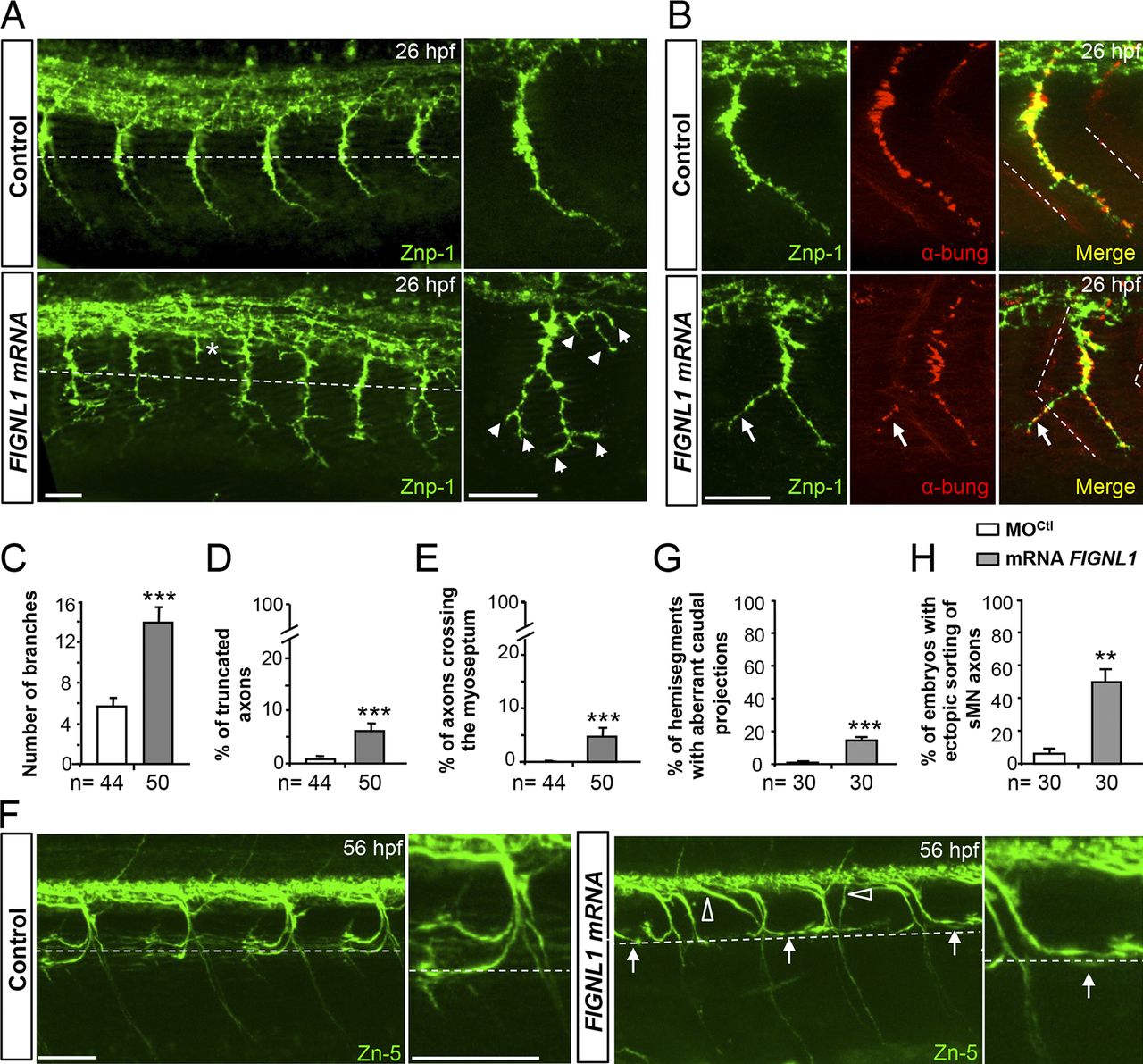
Fig. 5
FIGNL1 overexpression leads to spinal motor axon pathfinding defects. (A) Immunolabeling of pMN axons in 26-hpf control and human FIGNL1 mRNA–injected embryos with the use of Znp-1 antibody. Arrowheads and asterisk indicate hyperbranched or truncated pMN axons. (B) Analysis of neuromuscular junctions in 26-hpf control and FIGNL1 mRNA–injected embryos with the use of pre- (Znp-1, green) and postsynaptic markers. α-bung, α-bungarotoxin (red). Arrows indicate aberrant synapses between CaP axons and muscle fibers from the neighboring somite. (C) Mean number of branches. (D) Percentage of truncated axons. (E) Percentage of axons crossing the lateral myoseptum. (F) Immunolabeling of sMN axons with Zn-5 antibody in 56-hpf control and human FIGNL1 mRNA–injected larvae. Arrows show misguided rostral pMN–like sMNs. Open arrowheads indicate aberrant sMN axon exit points. (A, B, and F) Lateral views of the trunk; anterior is to the left. Right panels are higher magnifications of one SMN axon from the left panels. Dashed lines indicate the horizontal myotome (A and F) or lateral myosepta (B). Bars: (A and B) 25 µm; (F) 50 µm. (G) Percentage of hemisegments with aberrant caudal projections. (H) Percentage of embryos with ectopic sorting of sMN axons. The total number (n) of embryos from three independent experiments is indicated under the corresponding histogram bar. 24 hemisegments were analyzed per animal. **, P ≤ 0.01; ***, P ≤ 0.001; unpaired two-tailed t test. Error bars are SEM.