
Fig. 4
- ID
- ZDB-IMAGE-180723-15
- Antibodies
- Publication
- Fassier et al., 2018 - Motor axon navigation relies on Fidgetin-like 1-driven microtubule plus end dynamics
- All Figures
- Figures for Fassier et al., 2018
|
Fig. 4
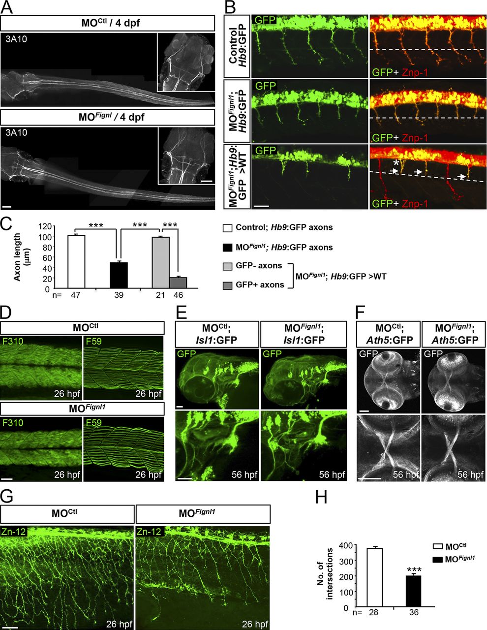
Fignl1 is mainly required for spinal motor axon targeting. (A) Immunolabeling of the Mauthner fibers in 4-dpf MOCtl (n = 17) and MOFignl1 (n = 19) larvae using the 3A10 antibody. Dorsal views of the larvae; anterior is to the left. Insets are higher magnifications of the head. (B) Immunostaining of SMN with Znp-1 and GFP antibodies in 30-hpf nontransplanted control (Hb9:GFP), morphant (MOFignl1; Hb9:GFP), and WT embryos transplanted with SMN precursors from MOFignl1 Tg(Hb9:GFP) embryos. The dashed line indicates the horizontal myoseptum. Arrows and asterisk show stalled or misrouted SMN axons, respectively. (C) Mean length of SMN axons. The number of axons analyzed is indicated under each histogram bar. (D) Immunostaining of slow and fast muscle fibers in 26-hpf MOCtl (n = 15) and MOFignl1 (n = 15) embryos using F59 and F310 antibodies. (E) Analysis of branchiomotor axons in 56-hpf MOCtl (n = 20) and MOFignl1 (n = 25) Tg(Islet1:GFP) larvae. Lateral views of the head; anterior is to the left. (F) Analysis of the optic nerve in 56-hpf MOCtl (n = 20) and MOFignl1(n = 20) Tg(Ath5:GFP) larvae. Ventral views of the head; anterior is to the left. Bottom panels are higher magnifications of top panels. (G) Immunostaining of mechanosensory RB neurons in 26-hpf MOCtl and MOFignl1 embryos. Skin innervation by RB axons is reduced in morphant embryos. (B, D, and G) Lateral views of the trunk; anterior is to the left. (H) Quantification of skin innervation using a stereological method (Rønn et al., 2000). The number of intersections between RB axons and a grid of 11 parallel lines spaced every 22 µm was estimated in 26-hpf MOCtl (n = 28) and MOFignl1 embryos (n = 36) from three independent experiments. ***, P ≤ 0.0001; unpaired two-tailed t test. Error bars are SEM. Bars: (A) 100 µm; (B) 25 µm; (D) 40 µm; (E and G) 50 µm; (F) 60 µm.