
Image
|
Figure Caption
Fig. 7
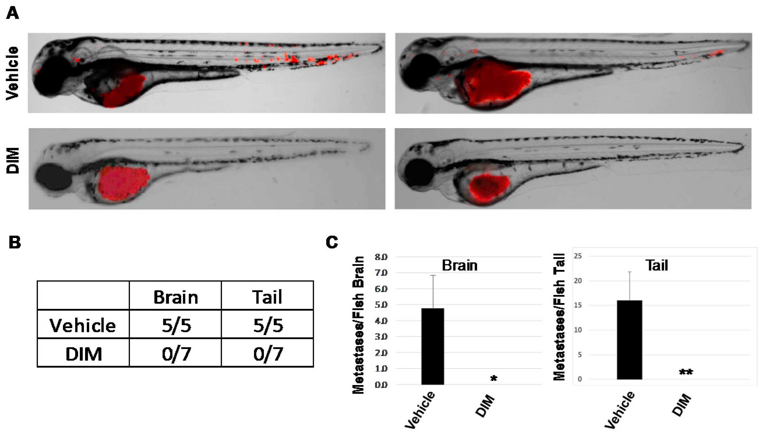
DIM inhibits metastasis in zebrafish larvae xenografts. RFP-HeLa cells were injected into the perivitiline space of two-day-old zebrafish larvae and larvae treated with vehicle (0.1% DMSO) or 1, 5, or 10 μM DIM (2–3 larvae/dose). Fish were imaged 24 h later at 8.5× magnification by confocal microscopy. (A) Left and right: two representative images from each group with 5–7 fish/group. (B) Number of fish with brain or tail metastases. (C) The number of metastases per fish were quantified and presented here as the average number of brain (left) or tail (right) metastases/fish + SE after treatment with vehicle (0.1% DMSO) or 1–10 μM DIM. * p < 0.05. ** p < 0.01.
Acknowledgments
This image is the copyrighted work of the attributed author or publisher, and
ZFIN has permission only to display this image to its users.
Additional permissions should be obtained from the applicable author or publisher of the image.
Full text @ Int. J. Mol. Sci.